. 2005 Feb;79(4):2309-24.
doi: 10.1128/JVI.79.4.2309-2324.2005.

The topology of bulges in the long stem of the flavivirus 3' stem-loop is a major determinant of RNA replication competence

Affiliations

The topology of bulges in the long stem of the flavivirus 3' stem-loop is a major determinant of RNA replication competence

Li Yu et al. J Virol. 2005 Feb.

Abstract

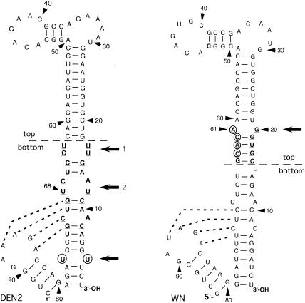
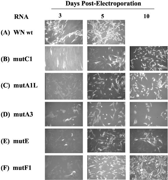
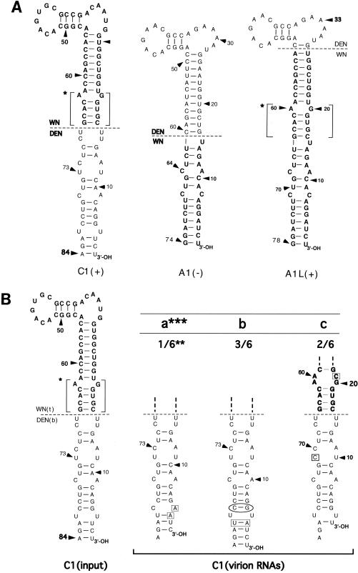
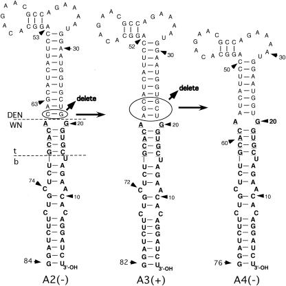
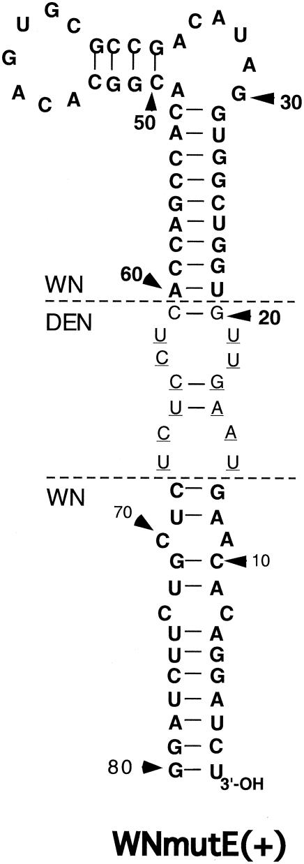
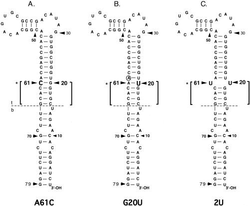
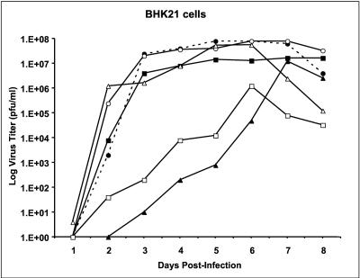
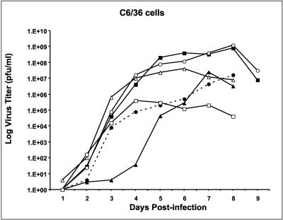
All flavivirus genomes contain a 3'terminal stem-loop secondary structure (3'SL) formed by the most downstream approximately 100 nucleotides (nt) of the viral RNA. The 3'SL is required for virus replication and has been shown to bind both virus-coded and cellular proteins. Results of the present study using an infectious DNA for WN virus strain 956 initially demonstrated that the dengue virus serotype 2 (DEN2) 3'SL nucleotide sequence could not substitute for that of the WN 3'SL to support WN genome replication. To determine what WN virus-specific 3'SL nucleotide sequences were required for WN virus replication, WN virus 3'SL nucleotide sequences were selectively deleted and replaced by analogous segments of the DEN2 3'SL nucleotide sequence such that the overall 3'SL secondary structure was not disrupted. Top and bottom portions of the WN virus 3'SL were defined according to previous studies (J. L. Blackwell and M. A. Brinton, J. Virol. 71:6433-6444, 1997; L. Zeng, L., B. Falgout, and L. Markoff, J. Virol. 72:7510-7522, 1998). A bulge in the top portion of the long stem of the WN 3'SL was essential for replication of mutant WN RNAs, and replication-defective RNAs failed to produce negative strands in transfected cells. Introduction of a second bulge into the bottom portion of the long stem of the wild-type WN 3'SL markedly enhanced the replication competence of WN virus in mosquito cells but had no effect on replication in mammalian cells. This second bulge was identified as a host cell-specific enhancer of flavivirus replication. Results suggested that bulges and their topological location within the long stem of the 3'SL are primary determinants of replication competence for flavivirus genomes.


Figures

FIG. 1.
FIG. 1.
The 3′-terminal 93-nt sequence of the DEN2 strain NGC 3′SL is shown on the left, and the 95-nt sequence of the WN virus strain 956 3′SL is shown on the right. Nucleotides are numbered in 3′-to-5′ direction from the 3′ terminus of genome RNA in both cases. Horizontal dashed lines indicate the chosen boundaries for the top and bottom portions of the respective 3′SLs, based on previous studies (3, 4, 42). Dotted lines between nucleotides of the respective small stem-loop structures and the long stems of both 3′SLs indicate putative pseudoknot structures (34). An 11-bp segment of the long stem in the DEN2 3′SL that was required for replication of mutant DEN2 RNAs containing DEN/WN virus chimeric 3′SL nucleotide sequences (DRS) is shown in boldface and underlined (42). Nucleotides comprising the putative major binding site for the TEF, eF1α, in the WN virus 3′SL, nt 64 to 61 (4), are circled. The 5-bp segment in the top part of the long stem in the WN virus 3′SL representing the TEF-binding domain is shown in boldface and underlined. Nucleotides U4-U76 which form a bulge in the bottom portion of the long stem of the DEN2 3′SL are circled. The loci of relevant bulges in the DRS (bulges 1 and 2) and the U4-U76 bulges in the DEN2 3′SL and the TEF-binding domain in the WN virus 3′SL are indicated by adjacent horizontal arrows. Arrowheads indicate nt numbers in the 3′-to-5′ direction.
FIG. 2.
FIG. 2.
Indirect IFAs after transfection of mutant WN virus RNAs. RNAs were derived by transfection of wt and mutant WN virus genome-length DNAs and used to transfect hamster kidney cells (BHK-21). On the days indicated, cells were replated on a chamber slide, and IFA was performed by standard methods with a polyclonal mouse anti-WN virus hyperimmune ascitic fluid on the days indicated. The nucleotide sequences of the 3′SLs in mutant C1, A1L, A3, E, and F1 RNAs are shown in Fig. 3A, 5, 6, and 8.
FIG. 3.
FIG. 3.
(A) Nucleotide sequences of mutant 3′SLs in WNmutC1, -A1, and -A1L RNAs, excluding that of the small stem and loop defined by wt WN virus nt 80 to 95 (Fig. 1) are shown. Nucleotides native to the DEN2 3′SL are shown in roman type. Nucleotides native to the wt WN virus 3′SL are shown in boldface. Horizontal dashed lines indicate the boundaries between WN virus and DEN2 nucleotide sequences, as labeled. The 5-bp TEF-binding domain in C1 RNA is indicated by brackets and an asterisk. (+), mutant RNA replicated efficiently after transfection of BHK (and Vero) cells, in that cells were 100% positive by IFA within 5 days posttransfection (see Fig. 2); (−), transfected cells remained negativeby IFA for 20 days posttransfection. (B) The nucleotide sequence of the long stem and loop at the 3′terminus of the WNmutC1 RNA transcribed from C1 mutant DNA and used to transfect cells is shown on the left (input). Spontaneous mutations of the C1 long stem and loop in genomes of replicating C1 virus are shown on the right (virion RNAs). The TEF-binding domain in C1 RNA is bracketed and marked by an asterisk. Boldface type indicates the WN 3′SL nucleotides. Roman type indicates DEN2 3′SL nucleotides. The horizontal dashed lines indicate the boundary between the top (t) and bottom (b) of the wt WN virus 3′SL (Fig. 1), which also constitutes the boundary between DEN2 (DEN) and WN virus nucleotide sequences in the C1 3′SL. The 3′SL in recovered C1 virus RNA was cloned, and six representative cloned DNAs were sequenced. ***a, b, and c indicate, respectively, each of the three spontaneous mutations of the C1 3′SL detected in C1 virion RNAs by this method. Top portions of the nucleotide sequences of C1 3′SL variants detected in replicating virus that did not deviate from that of input C1 RNA are indicated by the heavy vertical dashed lines. Nucleotides inserted or substituted into the C1 3′SL by spontaneous mutation are enclosed by a box or rectangle. Nucleotides spontaneously deleted from the C1 3′SL are enclosed in an oval. **, ratios of the respective mutant C1 nucleotide sequences in a total of six DNAs sequenced are indicated by fractions.
FIG. 4.
FIG. 4.
Nucleotide sequences of the long stem-loop structures of 3′SLs present in WNmutC2 (A) and WNmutA2 (B) RNAs are depicted in comparison to WNmutC1 and WNmutA1 3′SLs, respectively. Boldface type indicates nucleotides derived from the wt WN virus 3′SL sequence. Roman type, nucleotides derived from the wt DEN2 3′SL sequence. The 5-bp TEF-binding domain in C1 and A2 RNAs is indicated by brackets and an asterisk. Nucleotides included in the 11-bp ds segment previously shown to be required for DEN2 virus replication (42) are underlined. Arrows indicate that the TEF-binding domain was deleted from the C1 3′SL to generate the C2 3′SL. Similarly, arrows indicate that the TEF-binding domain was inserted into the A1 3′SL where shown in order to generate the A2 3′SL. Dashed lines delineate both the boundary between top and bottom portions of the WN virus 3′SL and the boundary between WN and DEN2 (DN) nucleotide sequences. (+), mutant RNA replicated efficiently after transfection of BHK (and Vero) cells compared to wt RNA; (−), transfected cells remained negative by IFA for 20 days posttransfection.
FIG. 5.
FIG. 5.
Derivation of the 3′SLs in WNmutA3 and WNmutA4 RNAs from the A2 3′SL nucleotide sequence is shown. Boldface type, nucleotides derived from the wt WN virus 3′SL nucleotide sequence. Roman type, nucleotides derived from the wt DEN2 3′SL nucleotide sequence. Arrows indicate that the G21-C65 base pair (enclosed by an oval) was deleted from the A2 3′SL to generate the A3 3′SL and that 3 bp (nt 21 to 23 and 61 to 63) (enclosed by an oval) were deleted from the A3 nucleotide sequence to generate the A4 3′SL nucleotide sequence. Horizontal dashed lines indicate, respectively, the boundary between DEN2 and WN virus nucleotide sequences in the context of the A2 3′SL and the top (t) and bottom (b) portions of the WN virus 3′SL, as defined in previous studies (3, 4, 42). Nucleotides comprising the TEF-binding domain (see text) are underlined. (+), mutant RNA replicated efficiently after transfection of BHK (and Vero) cells compared to wt RNA; (−), transfected cells remained negative by IFA for 20 days posttransfection.
FIG. 6.
FIG. 6.
The nucleotide sequence of the long stem-loop structure of the WNmutE 3′SL is depicted. Boldface type, nucleotides native to the wt WN virus 3′SL. Roman type, nucleotides native to the wt DEN2 3′SL. Horizontal dashed lines labeled WN and DEN indicate where a ds segment comprising wt WN virus nt 14 to 20 and 61 to 66, including the TEF-binding domain, was deleted and DEN2 nt 12 to 19 and 61 to 67 were inserted (Fig. 1). The inserted DEN2-specific nucleotides formed the major part of a ds segment shown in a previous study (42) to be required for replication of DEN2 RNAs containing chimeric DEN2-WN virus 3′SLs, and they are underlined.
FIG. 7.
FIG. 7.
Nucleotide sequences of the long stem and loop of 3′SLs present in A61C, G20U, and 2U RNAs are depicted. A horizontal line indicates the boundary between the top (t) and bottom (b) portions of the wt WN virus 3′SL (3, 4, 42). The TEF-binding domain in all three mutant nucleotide sequences is indicated by brackets and an asterisk to the left. For the A61C and G20U mutations, the loci of the substituted nucleotides are indicated in large, boldface type. For the 2U 3′SL, the U residues replacing A61 and G20, respectively, are shown in large boldface type. In the G20U nucleotide sequence, nucleotide A60, which spontaneously reverted to C in a portion of RNAs after transfection, is circled.
FIG. 8.
FIG. 8.
The nucleotide sequence of the long stem-loop structure in the WNmutF1 3′SL is depicted. Boldface type indicates nucleotides native to the wt WN 3′SL. Roman type indicates nucleotides native to the wt DEN2 3′SL. The horizontal dashed line indicates the boundary between DEN2 and WN virus 3′SL nucleotide sequences. The TEF-binding domain is indicated by brackets and an asterisk adjacent to the left-hand strand of the segment. Nucleotides forming the U-U bulge created in the WN sequence by substituting DEN2 nucleotides for Wnvirus nucleotides that comprise the lowermost 7 bp of the respective long stems (Fig. 1) are circled. (+), Mutant RNA replicated efficiently after transfection of BHK (and Vero) cells compared to wt RNA.
FIG. 9.
FIG. 9.
Replication of viable WN 3′SL mutant viruses in BHK cells. Plaque titers were determined for pools of viruses derived by transfection of BHK cells in Vero cells; these viruses were used to infect confluent monolayers of BHK cells at an MOI of 0.01. Plaque titers were determined for aliquots of the medium on infected cells on Vero cells on the days shown after infection. Results for wt WN virus strain 956 and WNmutA1L, WNmutA3, WNmutC1, WNmutE, and WNmutF1 viruses are shown. Solid circles and dashed line, wt WN virus; solid squares, WNmutC1 virus; solid triangles, WNmutE virus; open circles, WNmutF1 virus; open triangles, WNmutA3 virus; open squares, WNmutA1L virus.
FIG. 10.
FIG. 10.
Replication of viable WN 3′SL mutant viruses in C6/36 cells. Plaque titers were determined for pools of viruses derived by transfection of BHK cells in Vero cells; the viruses were used to infect confluent monolayers of C6/36 cells at an MOI of 0.01. Plaque titers were determined for aliquots of the medium on infected cells on Vero cells on the days shown after infection. Results for wt WN strain 956 and WNmutA1L, WNmutA3, WNmutC1, WNmutE, and WNmutF1 viruses are shown. Solid circles and dashed line, wt WN virus; solid squares, WNmutC1 virus; solid triangles, WNmutE virus; open circles, WNmutF1 virus; open triangles, WNmutA3 virus; open squares, WNmutA1L virus.
FIG. 11.
FIG. 11.
(A) Northern hybridization analysis of negative-strand WN RNAs in cells transfected by wt and lethal mutant RNAs. Total cellular RNAs were isolated from mock-transfected cells (lane 4) and from cells transfected with wt WN RNA (lane 3) or WNmutA1, WNmutA2, WNmutC2, and WN/DN-SL RNAs (lanes 5 to 8, respectively) 40 h p.e. RNAs were then electrophoresed on a denaturing 1.2% agarose gel for 4 h at 120V, transferred to a membrane, and hybridized to an in vitro-synthesized 32P-labeled ∼4.7-kb positive-sense ssDNA probe representing nt 1894 to 6777 of the WN virus genome. Full-length in vitro-synthesized WN negative-strand RNA (lane 1) and WN positive-strand RNA isolated from infectious virus (lane 2) served as controls to establish the specificity of the hybridization for WN virus negative-strand RNA. (B) After electrophoresis and prior to transblotting, the agarose gel was stained with ethidium bromide and photographed on a UV light box to visualize 18S and 28S rRNAs in preparations of total cellular RNA (lanes 3 to 8).

Similar articles

Cited by

References

    1. Bartel, D. P., M. L. Zapp, M. R. Green, and J. W. Szostak. 1991. HIV-1 Rev regulation involves recognition of non-Watson-Crick base pairs in the viral RNA. Cell 17:529-536. - PubMed
    1. Beasley, D. L., L. Li, M. T. Suderman, and A. D. Barrett. 2002. Mouse neuroinvasiveness phenotype of West Nile virus strains varies depending upon virus genotype. Virology 296:17-23. - PubMed
    1. Blackwell, J., and M. A. Brinton. 1996. BHK cell proteins that bind to the 3′ stem-loop structure of the West Nile virus genome RNA. J. Virol. 69:5650-5658. - PMC - PubMed
    1. Blackwell, J. L., and M. A. Brinton. 1997. Translation elongation factor-1 alpha interacts with the 3′ stem-loop region of West Nile virus genomic RNA. J. Virol. 71:6433-6444. - PMC - PubMed
    1. Brinton, M. A., A. V. Fernandez, and J. H. Dispoto. 1986. The 3′-nucleotides of flavivirus genomic RNA form a conserved secondary structure. Virology 153:113-121. - PubMed