. 2005 Feb;79(4):2528-40.
doi: 10.1128/JVI.79.4.2528-2540.2005.

Mutational analysis of narrow pores at the fivefold symmetry axes of adeno-associated virus type 2 capsids reveals a dual role in genome packaging and activation of phospholipase A2 activity

Affiliations

Mutational analysis of narrow pores at the fivefold symmetry axes of adeno-associated virus type 2 capsids reveals a dual role in genome packaging and activation of phospholipase A2 activity

Svenja Bleker et al. J Virol. 2005 Feb.

Abstract

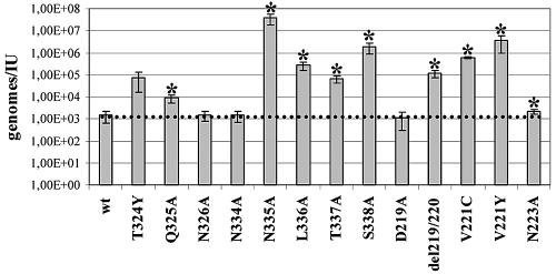
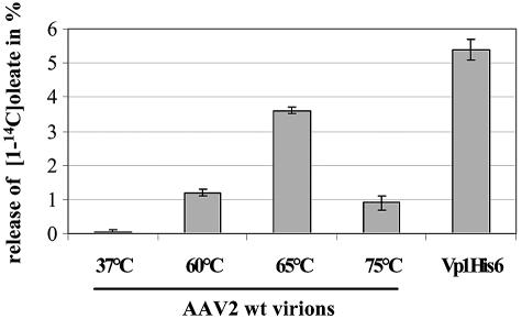
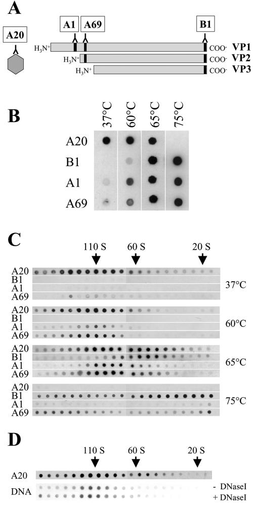
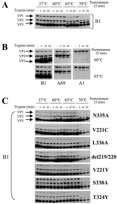
Adeno-associated virus type 2 (AAV2) capsids show 12 pores at the fivefold axes of symmetry. We mutated amino acids which constitute these pores to investigate possible functions of these structures within the AAV2 life cycle. Mutants with alterations in conserved residues were impaired mainly in genome packaging or infectivity, whereas few mutants were affected in capsid assembly. The packaging phenotype was characterized by increased capsid-per-genome ratios. Analysis of capsid-associated DNA versus encapsidated DNA revealed that this observation was due to reduced and not partial DNA encapsidation. Most mutants with impaired infectivity showed a decreased capability to expose their VP1 N termini. As a consequence, the activation of phospholipase A2 (PLA2) activity, which is essential for efficient infection, was affected on intact capsids. In a few mutants, the exposure of VP1 N termini and the development of PLA2 activity were associated with enhanced capsid instability, which is obviously also deleterious for virus infection. Therefore, PLA2 activity seems to be required on intact capsids for efficient infection. In conclusion, these results suggest that the pores at the fivefold axes function not only as portals for AAV2 single-stranded DNA packaging but also as channels for presentation of the PLA2 domain on AAV2 virions during infection.


Figures

FIG. 1.
FIG. 1.
Structures of pores at fivefold symmetry axes of AAV2 capsids. (A) Model of the AAV2 capsid (84); the arrow indicates the pore at a fivefold axis. (B and C) Backbone models of structures of fivefold symmetry-related VPs shown as a side view (B) and from the outside (C) of the capsid. Residues which form the pore are represented as space-filling models; different colors show that the pore is encircled by amino acids derived from each of the five subunits of the pentamer. (D) Schematic representation of the pore-forming amino acids (positions 217 to 223 and positions 322 to 338 of the VP sequence) shared by the three capsid proteins. (E) Alignment of pore-forming amino acids of eight known AAV serotypes; grey lettering indicates nonconserved amino acids.
FIG. 2.
FIG. 2.
Amino acids subjected to site-directed mutagenesis. (A) Residues of the AAV2 VP sequence chosen for site-directed mutagenesis (arrows denote the positions of the mutations); grey lettering indicates nonconserved amino acids. Charge-to-alanine mutations were made at positions 219, 223, 322, 325, 326, 334, 335, 336, 337, and 338; a tyrosine substitution at positions 221 and 324 and a cysteine substitution at position 221 were introduced. Additionally, a deletion of amino acids 219 and 220 (Δ), an insertion of two tyrosines after amino acid 221, and a triple substitution (amino acids 322, 323, and 324) were generated. (B and C) Localization of established mutations within pore structures at amino acids 217 and 223 (B) and amino acids 322 to 338 (C); a backbone model of a pentamer is shown as a side view. Amino acids which form the pore are represented as space-filling models; residues subjected to site-directed mutagenesis are shown in different colors. (D) Western blot analysis of 293T cells transfected with AAV2 wt or mutated genomic plasmids or Bluescript II (control) and subsequently superinfected with Ad5 (MOI, 2). AAV2 proteins were detected with monoclonal anti-Rep (303.9) or anti-VP (B1) antibodies. An extract from Ad5- and AAV2-coinfected HeLa cells was used as a marker (M).
FIG. 3.
FIG. 3.
Influence of mutation of pore-forming amino acids on genome packaging. (A) Virus supernatants obtained from 293T cells transfected with an AAV2 wt or mutated genomic plasmid (pTAV2.1: small Rep-deficient mutant) and superinfected with Ad5 (MOI, 2) were assayed for ELISA-based AAV2 capsid titers and dot blot-based genomic titers. Packaging was quantified as the ratio of capsids to genomes. Means ± standard deviations from at least four independent experiments are shown; an asterisk indicates a P value of <0.05 for a comparison with the wt value (indicated by the broken line). (B) Capsids were immunoprecipitated (IP) from frozen-thawed lysates obtained from 293T cells transfected with a plasmid harboring an AAV2 wt or mutated genome and superinfected with Ad5 (MOI, 2) by protein A-Sepharose-bound antibody A20. Control (C) precipitations were also performed with a non-AAV-related monoclonal antibody. Samples were digested (+) or not digested (−) with DNase I to discriminate between capsid-associated and packaged DNAs. Coprecipitated genomes were isolated and electrophoresed on neutral agarose gels. Southern blotting was performed, and genomes were detected with a rep-specific probe. A 4.7-kb fragment was used as a marker (top panels, M). A portion of each immunoprecipitate was tested by Western blot analysis for the level of capsid recovery; an extract from Ad5- and AAV2-coinfected HeLa cells was used as a marker (bottom panels, M).
FIG. 4.
FIG. 4.
Effect of mutation of pore-forming amino acids on infectivity. Virus supernatants obtained from 293T cells transfected with an AAV2 wt or mutated genomic plasmid and superinfected with Ad5 (MOI, 2) were assayed for dot blot-based genomic titers and dot blot-based infectious virus titers. Infectivity was quantified by calculating the ratios of genomes to infectious units (IU). This calculation corrects for the observed reduction in the packaging of some mutants. Means ± standard deviations from at least four independent experiments are shown; an asterisk indicates a P value of <0.05 for a comparison with the wt value (indicated by the broken line).
FIG. 5.
FIG. 5.
PLA2 activity of heat-treated AAV2 wt virions. AAV2 virions were exposed to various temperatures for 5 min and then incubated with [1-14C]oleate-labeled E. coli membranes. The release of [1-14C]oleate into supernatants was measured. As a positive control, the assay was performed with purified Vp1His6. Means ± standard deviations from at least three independent experiments are shown.
FIG. 6.
FIG. 6.
Exposure of VP1 N termini after heat treatment of AAV2 wt virions. (A) Localization of epitopes recognized by antibodies A20, A1, A69, and B1 in the native dot blot assay. (B) Native dot blot assay. AAV2 wt virions were exposed to various temperatures for 30 min and then reacted under nondenaturing conditions with antibodies A20, B1, A1, and A69. (C) AAV2 wt virions were incubated at various temperatures for 30 min and then loaded onto a continuous sucrose gradient (10 to 30% sucrose). Aliquots of each fraction were subjected to the native dot blot assay and probed with antibodies A20, B1, A1, and A69. (D) AAV2 wt virions were incubated at 65°C for 30 min and then loaded onto a continuous sucrose gradient (10 to 30% sucrose). Aliquots of each fraction were subjected to the native dot blot assay and detected with antibody A20. Another aliquot of each fraction was immunoprecipitated with antibody A20 and incubated with or without DNase I, and coprecipitated genomes were detected with a rep-specific probe. The 110S, 60S, and 20S positions were determined by using DNA-containing AAV2 particles, empty AAV2 particles, and thyroglobulin.
FIG. 7.
FIG. 7.
Influence of mutation of pore-forming amino acids on the accessibility of VP1 N termini after heat treatment. (A) Western blot analysis of AAV2 wt or mutant virus preparations with anti-VP monoclonal antibody B1. (B) AAV2 wt or mutant virions were incubated at various temperatures for 5 or 30 min, applied to nitrocellulose membranes, and then reacted under nondenaturing conditions with antibodies A20, B1, A1, and A69.
FIG. 8.
FIG. 8.
Accessibility of VP1 N termini to trypsin digestion. (A and B) AAV2 wt virions were incubated at various temperatures for 5 min, treated with trypsin for 5, 10, or 30 min, and then analyzed by Western blotting. VPs were detected with antibody B1 (A) or antibodies B1, A69, and A1 (B). (C) Mutant virions were incubated at various temperatures, treated with trypsin for 5, 10, or 30 min, and then analyzed by Western blotting. VPs were detected with antibody B1.
FIG. 9.
FIG. 9.
Effect of mutation of pore-forming amino acids on PLA2 activity. AAV2 wt and mutant virions were exposed to 60 and 65°C for 5 min and then incubated with [1-14C]oleate-labeled E. coli membranes. The release of [1-14C]oleate into supernatants was measured. Means ± standard deviations from at least three independent experiments are shown; an asterisk indicates a P value of <0.05 for a comparison with the wt value.

References

    1. Agbandje, M., R. McKenna, M. G. Rossmann, M. L. Strassheim, and C. R. Parrish. 1993. Structure determination of feline panleukopenia virus empty particles. Proteins 16:155-171. - PubMed
    1. Agbandje, M., S. Kajigaya, R. McKenna, N. S. Young, and M. G. Rossmann. 1994. The structure of human parvovirus B19 at 8 A resolution. Virology 203:106-115. - PubMed
    1. Agbandje-McKenna, M., A. L. Llamas-Saiz, F. Wang, P. Tattersall, and M. G. Rossmann. 1998. Functional implications of the structure of the murine parvovirus, minute virus of mice. Structure 6:1369-1381. - PubMed
    1. Atchison, R. W., B. C. Casto, and W. M. Hammon. 1965. Adeno-associated defective virus particles. Science 149:754-756. - PubMed
    1. Bartlett, J. S., R. Wilcher, and R. J. Samulski. 2000. Infectious entry pathway of adeno-associated virus and adeno-associated virus vectors. J. Virol. 74:2777-2785. - PMC - PubMed