

Kaposi's sarcoma-associated herpesvirus/human herpesvirus type 8-positive solid lymphomas: a tissue-based variant of primary effusion lymphoma
- PMID: 15681470
- PMCID: PMC1876263
- DOI: 10.1016/S1525-1578(10)60004-9
Kaposi's sarcoma-associated herpesvirus/human herpesvirus type 8-positive solid lymphomas: a tissue-based variant of primary effusion lymphoma
Abstract
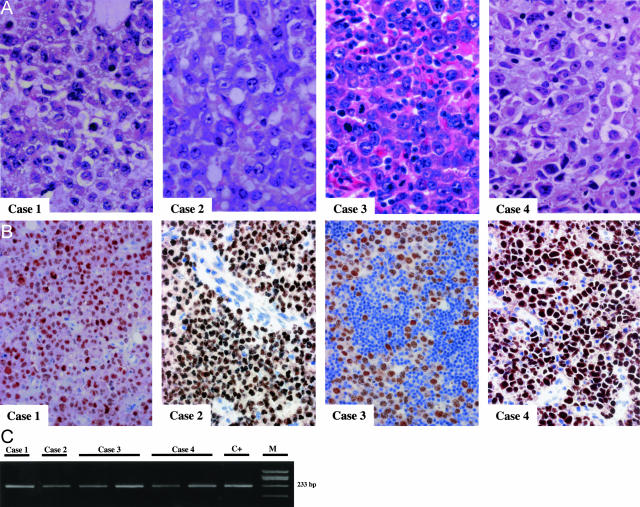
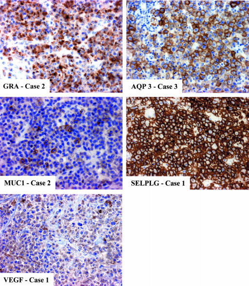
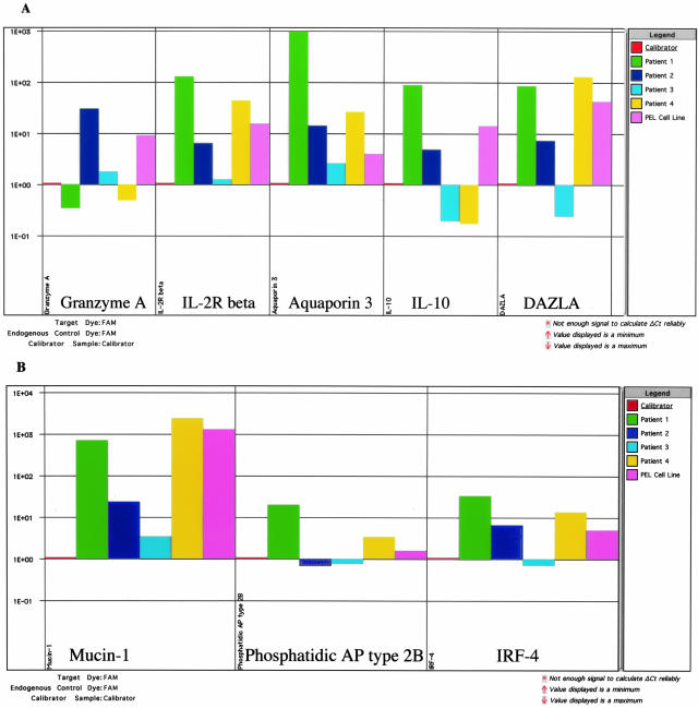
Kaposi's sarcoma-associated herpesvirus (KSHV), also termed human herpesvirus type 8, is consistently identified in Kaposi's sarcoma, primary effusion lymphoma (PEL), and multicentric Castleman's disease. Here we report four cases of KSHV-bearing solid lymphomas that occurred in AIDS patients (cases 1 to 3) and in a human immunodeficiency virus (HIV)-seronegative person (case 4). The patients presented extranodal masses in the abdomen (cases 1, 3, and 4) or skin (case 2), and nodal involvement, together with Kaposi's sarcoma (case 3). The gastrointestinal tract was involved in two patients (cases 1 and 3). The patients did not develop a lymphomatous effusion. KSHV was detected in the tumor cells of all cases by immunohistochemistry and by polymerase chain reaction. Epstein-Barr virus was detected in two of the HIV-related cases. All KSHV-positive solid lymphomas exhibited PEL-like cell morphology. To investigate the relationship of these disorders to PEL and to other AIDS-associated diffuse large cell lymphomas, KSHV-positive solid lymphomas were tested for the expression of a set of genes that were previously shown by gene profiling analysis to define PEL tumor cells. The results showed that expression of this set of genes in KSHV-positive lymphomas is similar to that of PEL but distinct from KSHV-negative AIDS-associated diffuse large cell lymphomas. Because pathobiological features of KSHV-positive solid lymphomas closely mimic those of PEL, our results suggest that KSHV-positive solid lymphomas should be considered as a tissue-based variant of classical PEL, irrespective of HIV status.
Figures
Similar articles
-
Human herpesvirus 8-associated solid lymphomas that occur in AIDS patients take anaplastic large cell morphology.Mod Pathol. 2000 Jan;13(1):77-85. doi: 10.1038/modpathol.3880012. Mod Pathol. 2000. PMID: 10658913
-
Kaposi's sarcoma-associated herpesvirus-like DNA sequences in AIDS-related body-cavity-based lymphomas.N Engl J Med. 1995 May 4;332(18):1186-91. doi: 10.1056/NEJM199505043321802. N Engl J Med. 1995. PMID: 7700311
-
Immunoblastic lymphoma in persons with AIDS-associated Kaposi's sarcoma: a role for Kaposi's sarcoma-associated herpesvirus.Mod Pathol. 2003 May;16(5):424-9. doi: 10.1097/01.MP.0000056629.62148.55. Mod Pathol. 2003. PMID: 12748248
-
Kaposi's sarcoma-associated herpesvirus: a lymphotropic human herpesvirus associated with Kaposi's sarcoma, primary effusion lymphoma, and multicentric Castleman's disease.Semin Diagn Pathol. 1997 Feb;14(1):54-66. Semin Diagn Pathol. 1997. PMID: 9044510 Review.
-
Kaposi sarcoma-associated herpesvirus (human herpesvirus type 8)-associated extracavitary lymphoma: Report of a case in an HIV-positive patient with simultaneous kaposi sarcoma and a review of the literature.Acta Haematol. 2010;123(4):237-41. doi: 10.1159/000314347. Epub 2010 May 19. Acta Haematol. 2010. PMID: 20484888 Review.
Cited by
-
KSHV/HHV-8 associated lymph node based lymphomas in HIV seronegative subjects. Report of two cases with anaplastic large cell morphology and plasmablastic immunophenotype.J Clin Pathol. 2005 Oct;58(10):1039-45. doi: 10.1136/jcp.2005.026542. J Clin Pathol. 2005. PMID: 16189148 Free PMC article.
-
The role of macrophages in the development and progression of AIDS-related non-Hodgkin lymphoma.J Leukoc Biol. 2010 Apr;87(4):627-32. doi: 10.1189/jlb.0809564. Epub 2009 Dec 30. J Leukoc Biol. 2010. PMID: 20042471 Free PMC article. Review.
-
Linking KSHV to human cancer.Curr Oncol Rep. 2005 Sep;7(5):349-56. doi: 10.1007/s11912-005-0061-6. Curr Oncol Rep. 2005. PMID: 16091195 Review.
-
Primary Effusion Lymphoma: Small Bowel Recurrence After Stem Cell Transplant.ACG Case Rep J. 2021 Nov 19;8(11):e00698. doi: 10.14309/crj.0000000000000698. eCollection 2021 Nov. ACG Case Rep J. 2021. PMID: 34820467 Free PMC article.
-
HHV-8-positive and EBV-positive intravascular lymphoma: an unusual presentation of extracavitary primary effusion lymphoma.Am J Surg Pathol. 2014 Mar;38(3):426-32. doi: 10.1097/PAS.0000000000000128. Am J Surg Pathol. 2014. PMID: 24525514 Free PMC article.
References
-
- Chang Y, Cesarman E, Pessin MS, Lee F, Culpepper J, Knowles DM, Moore PS. Identification of herpesvirus-like DNA sequences in AIDS-associated Kaposi’s sarcoma. Science. 1994;226:1865–1869. - PubMed
-
- Cesarman E, Knowles DM. The role of Kaposi’s sarcoma-associated herpesvirus (KSHV/HHV-8) in lymphoproliferative diseases. Semin Cancer Biol. 1999;9:165–174. - PubMed
-
- Cesarman E, Chang Y, Moore PS, Said JW, Knowles DM. Kaposi’s sarcoma-associated herpesvirus-like DNA sequences in AIDS-related body cavity-based lymphomas. N Engl J Med. 1995;332:1186–1191. - PubMed
-
- Soulier J, Grollet L, Oksenhendler E, Cacoub P, Cazals-Hatem D, Babinet P, d’Agay MF, Clauvel JP, Raphael M, Degos L. Kaposi’s sarcoma-associated herpesvirus-like DNA sequences in multicentric Castleman’s disease. Blood. 1995;86:1276–1280. - PubMed
-
- Dupin N, Diss TL, Kellam P, Tulliez M, Du MQ, Sicard D, Weiss RA, Isaacson PG, Boshoff C. HHV-8 is associated with a plasmablastic variant of Castleman disease that is linked to HHV-8-positive plasmablastic lymphoma. Blood. 2000;95:1406–1412. - PubMed
Publication types
MeSH terms
Grants and funding
LinkOut - more resources
Full Text Sources
Medical
