. 2005 Feb 15;19(4):472-88.
doi: 10.1101/gad.1267105. Epub 2005 Jan 28.

The kinase haspin is required for mitotic histone H3 Thr 3 phosphorylation and normal metaphase chromosome alignment

Affiliations

The kinase haspin is required for mitotic histone H3 Thr 3 phosphorylation and normal metaphase chromosome alignment

Jun Dai et al. Genes Dev. .

Abstract

Post-translational modifications of conserved N-terminal tail residues in histones regulate many aspects of chromosome activity. Thr 3 of histone H3 is highly conserved, but the significance of its phosphorylation is unclear, and the identity of the corresponding kinase unknown. Immunostaining with phospho-specific antibodies in mammalian cells reveals mitotic phosphorylation of H3 Thr 3 in prophase and its dephosphorylation during anaphase. Furthermore we find that haspin, a member of a distinctive group of protein kinases present in diverse eukaryotes, phosphorylates H3 at Thr 3 in vitro. Importantly, depletion of haspin by RNA interference reveals that this kinase is required for H3 Thr 3 phosphorylation in mitotic cells. In addition to its chromosomal association, haspin is found at the centrosomes and spindle during mitosis. Haspin RNA interference causes misalignment of metaphase chromosomes, and overexpression delays progression through early mitosis. This work reveals a new kinase involved in composing the histone code and adds haspin to the select group of kinases that integrate regulation of chromosome and spindle function during mitosis and meiosis.


Figures

Figure 1.
Figure 1.
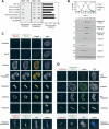
Haspin is a nuclear protein during interphase and associates with the chromosomes and spindle apparatus during mitosis. (A) Interphase HeLa cells transiently transfected with a vector encoding myc-haspin were analyzed by immunofluorescent staining with anti-myc-FITC (green) and anti-nucleophosmin/B23 followed by anti-goat IgG-Cy3 antibodies (red). DNA was visualized with Hoechst 33342. The arrowhead indicates a nontransfected cell. (B) Live cell confocal fluorescence microscopy of HeLa cells stably transfected with a vector encoding EGFP-haspin under a doxycycline-inducible promoter and treated with doxycycline for 1 d. DNA was visualized with the cell-permeable dye DRAQ5 (blue). Images on the left are projections of confocal image stacks and those on the right show individual confocal sections from a single metaphase, anaphase, and telophase cell. Arrows indicate the presence of haspin at the centrosomes, and arrowheads indicate haspin associated with the spindle. (C) Metaphase chromosome spreads from myc-haspin transfected HeLa cells were fixed and stained with anti-myc-FITC (green) and DNA was visualized with DRAQ5 (blue). (D) HeLa cells treated as in B were analyzed by live cell epifluorescence video microscopy at 37°C. Selected images of EGFP fluorescence (green) are shown superimposed on corresponding bright-field images. The entire series is available as a Supplementary Movie. (E) Confocal immunofluorescence microscopy of a metaphase HeLa cell expressing EGFP-haspin (green), treated with 0.2% Triton X-100 in PHEM buffer followed by 3% formaldehyde. DNA was visualized with DRAQ5 (blue) and mouse anti-α-tubulin staining with anti-mouse IgG-Cy3 (red). Arrows indicate the presence of haspin but not DNA at the spindle poles.
Figure 2.
Figure 2.
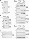
Haspin is a histone H3 kinase in vitro. (A) HEK293 cells were transiently transfected with a vector encoding human haspin or vector alone. Fractions enriched for cytoplasmic (C) and nuclear (N) components were obtained by hypotonic lysis and analyzed by immunoblotting with affinity-purified rabbit antihaspin 329–44 antibodies. Appropriate fractionation was verified by immunoblotting for the endoplasmic reticulum protein calnexin and the nuclear protein histone H3. (B) HEK293 cells were transiently transfected with vectors encoding myc-haspin or myc-haspin-KD or with vector alone. Rabbit antihaspin immunoprecipitates from lysed cells were subjected to in vitro kinase reactions with γ32P-ATP as described in Materials and Methods. That equivalent amounts of myc-haspin and myc-haspin-KD were present in the immunoprecipitates was verified by immunoblotting with rabbit antihaspin antibodies. (C) In vitro kinase reactions of myc-haspin or myc-haspin-KD with or without the addition of purified calf thymus histones as an exogenous substrate were carried out as described in B. The positions of the histones in the gel were determined by Coomassie blue staining. (D) In vitro kinase reactions of rabbit antihaspin immunoprecipitates from myc-haspin- or vector-alone-transfected HeLa cells using intact polynucleosomes (with or without the linker histone H1) as exogenous substrates were carried out as described in B. (E) In vitro kinase reactions of rabbit anti-haspin immunoprecipitates from myc-haspin- or vector-alone-transfected HEK293 cells with recombinant Xenopus histones purified from E. coli as exogenous substrates were carried out as described in B. (gH3) Tailless histone H3.
Figure 5.
Figure 5.
Haspin is phosphorylated during mitosis, and its overexpression delays mitotic progression. (A) HeLa Tet-On/myc-haspin- or vector-alone-stably-transfected cells were induced with 1 μg/mL doxycycline for the times indicated, and myc-haspin expression was assessed by rabbit antihaspin immunoblotting. (B) HeLa Tet-On/myc-haspin- or vector-alone-stably-transfected cells were induced with 1 μg/mL doxycycline for 24 h before synchronization at G1/S and analysis as described for Figure 4B. (C) Synchronized HeLa Tet-On/myc-haspin cells as shown in B were lysed and analyzed by immunoblotting and haspin in vitro kinase assay with γ32P-ATP using H3–GST or H3-T3A–GST as a control. (D) Synchronized HeLa Tet-On/vector cells as shown in B were analyzed as described in C.(E) Induced HeLa Tet-On/myc-haspin cells were fractionated into looselyadherent mitotic (mitosis) and adherent interphase-enriched (interphase) populations or treated with colcemid prior to lysis. Rabbit antihaspin or rabbit IgG control immunoprecipitates were analyzed by rabbit antihaspin immunoblotting. (F) Induced HeLa Tet-On/myc-haspin cells were treated colcemid for 12 h prior to release. Whole-cell lysates made at the times indicated were analyzed by antihaspin immunoblotting. (G) Induced HeLa Tet-On/myc-haspin cells were treated colcemid for 12 h or untreated prior to cell lysis. Antihaspin immunoprecipitates were incubated with or without λ phosphatase as described in Materials and Methods and analyzed by antihaspin immunoblotting.
Figure 3.
Figure 3.
Haspin associates with histone H3 and phosphorylates it at Thr 3. (A) HeLa cells were transfected with vectors encoding myc-haspin or vector alone. Immunoprecipitates from cell lysates with rabbit antihaspin antibodies (haspin) or with nonimmune rabbit IgG (neg) were subjected to in vitro kinase assays with γ32P-ATP and histone tail–GST proteins as exogenous substrates. Equivalent loading of the GST proteins was verified by Coomassie blue staining. (B) Lysates of HEK293 cells transiently transfected with a vector encoding myc-haspin were incubated with glutathione-Sepharose beads coated with histone tail–GST proteins. After washing, the binding of myc-haspin, cyclin A, and PCNA to the beads was assessed by immunoblotting. The lane marked lysate was loaded with 1/20 volume of the input lysate, and equivalent loading of the GST proteins was verified by Coomassie blue staining of the blot. (C) Peptides representing histone H3 amino acids 1–8 either without [H3(1–8)] or with phosphorylated Thr 3 [H3(1–8)pT3] or peptides containing phosphorylated Thr 11 [H3(9–16)pT11] or Thr 22 [H3(20–27)pT22] were immobilized by slot blotting and probed with rabbit anti-phospho-histone H3 (Thr-3) antibody B8634. Similar results were obtained with the commercial polyclonal anti-phospho-histone H3 (Thr-3) antibody (Upstate), although some lower cross-reactivity with the phospho-Thr 22 peptide was observed in this case. (D) Purified recombinant wild-type 6His-haspin kinase domain (kinase) or 6His-kinase containing the mutation K511A (kinase-KD) was examined by SDS-PAGE and Coomassie blue staining. (E) In vitro kinase reactions were carried out using 15 ng recombinant haspin 6His-kinase domain (kinase) or 6His-kinase-KD (kinase-KD) with 0.5 μg purified recombinant H3, H3–GST, H3-T3A–GST, and GST alone as substrates. The products were analyzed by immunoblotting with rabbit anti-phospho-H3 (Thr-3), followed by staining with Coomassie blue to verify similar loading of substrate proteins.
Figure 4.
Figure 4.
Histone H3 is phosphorylated at Thr 3 during mitosis. (A) Biotinylated peptides representing histone H3 residues 1–21 with various modifications were phosphorylated at Thr 3 in vitro using the haspin 6His-kinase domain (haspin) or were mock-treated in the absence of the kinase (none). (Right panel) The extent of phosphorylation was assessed by incorporation of radiolabeled phosphate from γ32P-ATP. Peptides immobilized by slot blotting were probed with rabbit anti-phospho-histone H3 (Thr-3) (left panel), and equivalent peptide loading was confirmed by streptavidin binding (center panel). Similar results were obtained with both anti-phospho-H3 (Thr-3) antibodies. (B) HeLa cells were synchronized at G1/S by double thymidine block. At various times following removal of thymidine, progression through the cell cycle was followed by propidium iodide staining for DNA content (G1 and G2/M) and MPM-2-FSE staining to enumerate mitotic cells (M), and cell lysates were analyzed by immunoblotting as indicated. (C) HeLa cells or metaphase spreads were fixed and stained with mouse anti-phospho-H3 (Ser-10) followed by anti-mouse IgG-Cy3 (red) and rabbit anti-phospho-H3 (Thr-3) followed by anti-rabbit IgG-Alexa488 (green). Cells were examined by confocal fluorescence microscopy, and the stages of mitosis were determined by the distinctive pattern of DNA visualized by DRAQ5 staining. (D) HeLa cells or metaphase spreads were stained as described in C with human centromeric autoantibodies followed by antihuman IgG-Cy3 (red) and rabbit anti-phospho-H3 (Thr-3) followed by anti-rabbit IgG-Alexa488 (green).
Figure 6.
Figure 6.
Endogenous haspin is required for phosphorylation of histone H3 on Thr 3. (A) Immunoprecipitates with rabbit antihaspin or rabbit IgG negative control (neg) antibodies from HeLa cell lysates were subjected to in vitro kinase assays with γ32P-ATP and H3–GST or H3-T3A–GST as substrates. Coomassie blue staining was used to confirm equivalent levels of GST proteins. (B) Antihaspin and anti-aurora B immunoprecipitates from HeLa cell lysates were subjected to in vitro kinase reactions with γ32P-ATP and H3–GST, H3-T3A–GST, or H3-S10A–GST proteins as substrates. Coomassie blue staining was used to confirm equivalent levels of GST proteins. (C) The synchronized HeLa Tet-On/vector cells shown in Figure 5B were analyzed by haspin in vitro kinase assay described for Figure 5C. Note that this is a longer exposure of the autoradiogram shown in Figure 5D, carried out in order to visualize endogenous haspin activity. Similar results were obtained with HeLa cells (data not shown). (D) HeLa cells were transfected with haspin siRNA (Ambion ID #1093), control siRNA (Ambion 4611), or no siRNA. Approximately 30 h after transfection, the cells were incubated with nocodazole for 16 h (mitotic) or left untreated (asynchronous) prior to lysis. Antihaspin and anti-aurora B immunoprecipitates were subjected to in vitro kinase reactions with γ32P-ATP and H3–GST, H3-T3A–GST, or H3-S10A–GST as substrates, and lysates were immunoblotted with anti-phospho-H3 (Thr-3) or anti-phospho-H3 (Ser-10) antibodies. The anti-phospho-H3 (Thr-3) blot was stripped and reprobed with antihistone H3 antibodies. The increased haspin activity in nocodazole-treated cells was not a reproducible finding, as shown in B and C. (E) NIH3T3 cells were transfected with mouse haspin (Ambion ID 67120) or negative control (Ambion 4611) siRNAs and analyzed as described for D.
Figure 7.
Figure 7.
Depletion of haspin causes a failure of chromosome congression. (A) U2OS cells transfected with haspin siRNA (Ambion ID #1093) or control siRNA (Ambion #4613) were fixed and stained with DRAQ5 to visualize DNA and with human centromeric autoantibodies followed by antihuman IgG-Cy3 (red), mouse anti-α-tubulin mAb followed by anti-mouse IgG-Alexa488 (green), and rabbit anti-phospho-H3 (Thr-3) followed by anti-rabbit IgG-Alexa488 (green) or anti-rabbit IgG-Cy3 (red) as indicated. (B) U2OS cells were transfected with haspin siRNA A (Ambion ID #1093), haspin siRNA B (Dharmacon SMARTpool), control siRNA A (Ambion #4613), or control siRNA B (Dharmacon SMARTpool), or without siRNA. After 48 h, the cells were fixed and stained with propidium iodide, and ∼3000 cells were counted on each of three coverslips for each condition and classified as interphase, prophase, prometaphase, metaphase, anaphase/telophase, or partial metaphase (a metaphase plate and three or more unaligned chromosomes; a subset of prometaphase) by the distinctive pattern of DNA staining. The percent of mitotic cells in each phase is shown. The increase in prometaphase and partial metaphase cells and the decrease in anaphase/telophase cells have p < 0.0001 by two-tailed Student's t-test when comparing haspin siRNA A and B with control siRNA A, B, and no siRNA combined. (C) Haspin siRNA transfected U2OS cells in prometaphase and metaphase from B stained with rabbit anti-phospho-H3 (Thr-3) followed by anti-rabbit IgG-Alexa488 were further classified as containing low or high levels of phosphorylated H3 Thr 3 (pT3). The percentage of these cells that were in prometaphase, metaphase, or partial metaphase is shown.

Similar articles

Cited by

References

    1. Adams R.R., Maiato, H., Earnshaw, W.C., and Carmena, M. 2001. Essential roles of Drosophila inner centromere protein (INCENP) and aurora B in histone H3 phosphorylation, metaphase chromosome alignment, kinetochore disjunction, and chromosome segregation. J. Cell. Biol. 153: 865–880. - PMC - PubMed
    1. Aihara H., Nakagawa, T., Yasui, K., Ohta, T., Hirose, S., Dhomae, N., Takio, K., Kaneko, M., Takeshima, Y., Muramatsu, M., et al. 2004. Nucleosomal histone kinase-1 phosphorylates H2A Thr 119 during mitosis in the early Drosophila embryo. Genes & Dev. 18: 877–888. - PMC - PubMed
    1. Andrews P.D., Knatko, E., Moore, W.J., and Swedlow, J.R. 2003. Mitotic mechanics: The auroras come into view. Curr. Opin. Cell. Biol. 15: 672–683. - PubMed
    1. Barr F.A., Sillje, H.H., and Nigg, E.A. 2004. Polo-like kinases and the orchestration of cell division. Nat. Rev. Mol. Cell. Biol. 5: 429–440. - PubMed
    1. Bischoff J.R., Anderson, L., Zhu, Y., Mossie, K., Ng, L., Souza, B., Schryver, B., Flanagan, P., Clairvoyant, F., Ginther, C., et al. 1998. A homologue of Drosophila aurora kinase is oncogenic and amplified in human colorectal cancers. EMBO J. 17: 3052–3065. - PMC - PubMed

Publication types