

doi: 10.1101/gad.326705.
Epub 2005 Jan 28.
Frat is dispensable for canonical Wnt signaling in mammals
Affiliations
- PMID: 15681612
- PMCID: PMC548942
- DOI: 10.1101/gad.326705
Item in Clipboard
Frat is dispensable for canonical Wnt signaling in mammals
Genes Dev.
.
Abstract
Wnt-signal transduction through beta-catenin is thought to require the inhibition of GSK3 by Frat/GBP. To investigate the role of Frat in mammalian development, we have generated mice with targeted mutations in all three murine Frat homologs. We show that Frat is normally expressed at sites of active Wnt signaling. Surprisingly, Frat-deficient mice do not display gross abnormalities. Moreover, canonical Wnt signaling in primary cells is unaffected by the loss of Frat. These studies show that Frat is not an essential component of the canonical Wnt pathway in higher organisms, despite the strict requirement of Frat/GBP for maternal Wnt signaling in Xenopus.
Figures
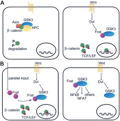
Model for the physiological role of Frat. (A) Current model of canonical Wnt-signal transduction. See text for details. (B) Several models could explain the observation that Frat is not critically required for canonical Wnt signaling in mammals. (Left) Frat might be induced under specific conditions or it might feed into the canonical Wnt pathway from a parallel route independent from upstream Wnt signals. (Right) Alternatively, Frat might be involved in any of the numerous other GSK3-dependent cellular activities, such as the NFκB and NFAT signaling pathways.
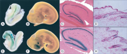
Frat1 and Frat2 show an identical expression pattern in neural and epithelial tissues of developing and adult mice. Comparison of lacZ activity in Frat1+/lacZ (A–D) and Frat2+/lacZ (E–H) mice reveals overlapping expression between the two homologs. (A,E) Embryonic day 8.5 (E8.5) embryo. (B,F) E10.5 embryo. (C,G) Adult hippocampus. (D,H) Adult skin.
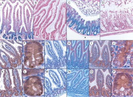
Loss of Frat does not affect the intestinal Wnt gradient. (A–D) LacZ staining on duodenum (A,B) and colon (C,D) shows that Frat2 expression is restricted to intestinal crypt cells. (A,C) Frat2+/lacZ. (B,D) Littermate control. (E–P) The expression of different Wnt-related markers is unchanged in Frat-TKO intestine. (E–J) Frat-TKO. (K–P) Littermate control. (E,K) β-Catenin, 40×. F and L are close-ups of crypts depicted in E and K, respectively. (G,M) Ki67, 40×.(H,N) EphrinB1, 40×.(I,O) EphB2, 40×. J and P are close-ups of crypts depicted in I and O, respectively.
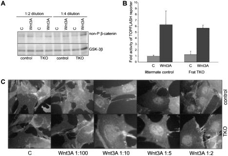
Sensitivity to Wnt ligand and the β-catenin/TCF response are unimpaired in Frat-TKO cells. TKO and littermate control MEFs were stimulated with control L-cell (lanes labeled C) or Wnt3A-CM (Wnt3A). Activation of canonical Wnt signaling was determined by Western blot analysis of unphosphorylated β-catenin (A), TOPFLASH luciferase reporter activity (shown are the averages of two experiments on independent MEF isolates, each performed in triplicate) (B), and immunofluorescence to visualize the nuclear accumulation of β-catenin following titration of different dilutions of Wnt3A-CM (ranging from 1:100 to 1:2) (C).
Similar articles
-
Frat oncoproteins act at the crossroad of canonical and noncanonical Wnt-signaling pathways.Oncogene. 2010 Jan 7;29(1):93-104. doi: 10.1038/onc.2009.310. Epub 2009 Oct 5. Oncogene. 2010. PMID: 19802005
-
Re-evaluating the role of Frat in Wnt-signal transduction.Cell Cycle. 2005 Aug;4(8):1065-72. Epub 2005 Aug 1. Cell Cycle. 2005. PMID: 16082208 Review.
-
TOPGAL mice show that the canonical Wnt signaling pathway is active during bone development and growth and is activated by mechanical loading in vitro.J Bone Miner Res. 2005 Jul;20(7):1103-13. doi: 10.1359/JBMR.050210. Epub 2005 Feb 14. J Bone Miner Res. 2005. PMID: 15940363
-
Dkk1-induced inhibition of Wnt signaling in osteoblast differentiation is an underlying mechanism of bone loss in multiple myeloma.Bone. 2008 Apr;42(4):669-80. doi: 10.1016/j.bone.2007.12.006. Epub 2007 Dec 27. Bone. 2008. PMID: 18294945
-
WNT signaling pathway and stem cell signaling network.Clin Cancer Res. 2007 Jul 15;13(14):4042-5. doi: 10.1158/1078-0432.CCR-06-2316. Clin Cancer Res. 2007. PMID: 17634527 Review.
Cited by
-
Overexpression of Frat1 correlates with malignant phenotype and advanced stage in human non-small cell lung cancer.Virchows Arch. 2011 Sep;459(3):255-63. doi: 10.1007/s00428-011-1135-5. Epub 2011 Aug 5. Virchows Arch. 2011. PMID: 21818639
-
What does genetics tell us about imprinting and the placenta connection?Cell Mol Life Sci. 2015 Jan;72(1):51-72. doi: 10.1007/s00018-014-1714-0. Epub 2014 Sep 7. Cell Mol Life Sci. 2015. PMID: 25194419 Free PMC article. Review.
-
The clinical pathological significance of FRAT1 and ROR2 expression in cartilage tumors.Clin Transl Oncol. 2015 Jun;17(6):438-45. doi: 10.1007/s12094-014-1254-y. Epub 2014 Nov 12. Clin Transl Oncol. 2015. PMID: 25387569
-
Significant Genes Associated with Mortality and Disease Progression in Grade II and III Glioma.Biomedicines. 2024 Apr 12;12(4):858. doi: 10.3390/biomedicines12040858. Biomedicines. 2024. PMID: 38672212 Free PMC article.
-
A novel Axin2 knock-in mouse model for visualization and lineage tracing of WNT/CTNNB1 responsive cells.Genesis. 2020 Sep;58(9):e23387. doi: 10.1002/dvg.23387. Epub 2020 Jul 9. Genesis. 2020. PMID: 32643876 Free PMC article.
References
-
- Anderson K.V. and Ingham, P.W. 2003. The transformation of the model organism: A decade of developmental genetics. Nat. Genet. 33 Suppl: 285–293. - PubMed
-
- Batlle E., Henderson, J.T., Beghtel, H., van den Born, M.M., Sancho, E., Huls, G., Meeldijk, J., Robertson, J., van de Wetering, M., Pawson, T., et al. 2002. β-Catenin and TCF mediate cell positioning in the intestinal epithelium by controlling the expression of EphB/ephrinB. Cell 111: 251–263. - PubMed
-
- Bax B., Carter, P.S., Lewis, C., Guy, A.R., Bridges, A., Tanner, R., Pettman, G., Mannix, C., Culbert, A.A., Brown, M.J., et al. 2001. The structure of phosphorylated GSK-3β complexed with a peptide, FRATtide, that inhibits β-catenin phosphorylation. Structure (Camb) 9: 1143–1152. - PubMed
Publication types
MeSH terms
Substances
LinkOut - more resources
Full Text Sources
Molecular Biology Databases
Research Materials