Surface behavior and lipid interaction of Alzheimer beta-amyloid peptide 1-42: a membrane-disrupting peptide
- PMID: 15681641
- PMCID: PMC1305366
- DOI: 10.1529/biophysj.104.055582
Surface behavior and lipid interaction of Alzheimer beta-amyloid peptide 1-42: a membrane-disrupting peptide
Abstract
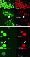
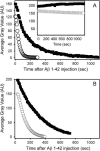
Amyloid aggregates, found in patients that suffer from Alzheimer's disease, are composed of fibril-forming peptides in a beta-sheet conformation. One of the most abundant components in amyloid aggregates is the beta-amyloid peptide 1-42 (Abeta 1-42). Membrane alterations may proceed to cell death by either an oxidative stress mechanism, caused by the peptide and synergized by transition metal ions, or through formation of ion channels by peptide interfacial self-aggregation. Here we demonstrate that Langmuir films of Abeta 1-42, either in pure form or mixed with lipids, develop stable monomolecular arrays with a high surface stability. By using micropipette aspiration technique and confocal microscopy we show that Abeta 1-42 induces a strong membrane destabilization in giant unilamellar vesicles composed of palmitoyloleoyl-phosphatidylcholine, sphingomyelin, and cholesterol, lowering the critical tension of vesicle rupture. Additionally, Abeta 1-42 triggers the induction of a sequential leakage of low- and high-molecular-weight markers trapped inside the giant unilamellar vesicles, but preserving the vesicle shape. Consequently, the Abeta 1-42 sequence confers particular molecular properties to the peptide that, in turn, influence supramolecular properties associated to membranes that may result in toxicity, including: 1), an ability of the peptide to strongly associate with the membrane; 2), a reduction of lateral membrane cohesive forces; and 3), a capacity to break the transbilayer gradient and puncture sealed vesicles.
Figures


Similar articles
-
Molecular interactions of Alzheimer amyloid-β oligomers with neutral and negatively charged lipid bilayers.Phys Chem Chem Phys. 2013 Jun 21;15(23):8878-89. doi: 10.1039/c3cp44448a. Epub 2013 Mar 14. Phys Chem Chem Phys. 2013. PMID: 23493873 Free PMC article.
-
How cholesterol constrains glycolipid conformation for optimal recognition of Alzheimer's beta amyloid peptide (Abeta1-40).PLoS One. 2010 Feb 5;5(2):e9079. doi: 10.1371/journal.pone.0009079. PLoS One. 2010. PMID: 20140095 Free PMC article.
-
Effects of membrane interaction and aggregation of amyloid β-peptide on lipid mobility and membrane domain structure.Phys Chem Chem Phys. 2013 Jun 21;15(23):8929-39. doi: 10.1039/c3cp44517h. Epub 2013 Mar 21. Phys Chem Chem Phys. 2013. PMID: 23515399
-
Amyloid-β Interactions with Lipid Rafts in Biomimetic Systems: A Review of Laboratory Methods.Methods Mol Biol. 2021;2187:47-86. doi: 10.1007/978-1-0716-0814-2_4. Methods Mol Biol. 2021. PMID: 32770501 Review.
-
Aggregation Behavior of Amyloid Beta Peptide Depends Upon the Membrane Lipid Composition.J Membr Biol. 2024 Aug;257(3-4):151-164. doi: 10.1007/s00232-024-00314-3. Epub 2024 Jun 18. J Membr Biol. 2024. PMID: 38888760 Review.
Cited by
-
Monosialoganglioside-GM1 triggers binding of the amyloid-protein salmon calcitonin to a Langmuir membrane model mimicking the occurrence of lipid-rafts.Biochem Biophys Rep. 2016 Oct 15;8:365-375. doi: 10.1016/j.bbrep.2016.10.005. eCollection 2016 Dec. Biochem Biophys Rep. 2016. PMID: 28955978 Free PMC article.
-
Mechanical properties of the brain: Focus on the essential role of Piezo1-mediated mechanotransduction in the CNS.Brain Behav. 2023 Sep;13(9):e3136. doi: 10.1002/brb3.3136. Epub 2023 Jun 27. Brain Behav. 2023. PMID: 37366640 Free PMC article. Review.
-
CLIC1 function is required for beta-amyloid-induced generation of reactive oxygen species by microglia.J Neurosci. 2008 Nov 5;28(45):11488-99. doi: 10.1523/JNEUROSCI.2431-08.2008. J Neurosci. 2008. PMID: 18987185 Free PMC article.
-
AβP1-42 incorporation and channel formation in planar lipid membranes: the role of cholesterol and its oxidation products.J Bioenerg Biomembr. 2013 Aug;45(4):369-81. doi: 10.1007/s10863-013-9513-0. Epub 2013 Apr 26. J Bioenerg Biomembr. 2013. PMID: 23620083
-
Absence of fluid-ordered/fluid-disordered phase coexistence in ceramide/POPC mixtures containing cholesterol.Biophys J. 2006 Jun 15;90(12):4437-51. doi: 10.1529/biophysj.105.077107. Epub 2006 Mar 24. Biophys J. 2006. PMID: 16565051 Free PMC article.
References
-
- Ambroggio, E. E., F. Separovic, J. Bowie, and G. D. Fidelio. 2004. Surface behaviour and peptide-lipid interactions of the antibiotic peptides, Maculatin and Citropin. Biochim. Biophys. Acta. 1664:31–37. - PubMed
-
- Angelova, M. I., and D. S. Dimitrov. 1986. Liposome electroformation. Faraday Discuss. Chem. Soc. 81:303–311.
-
- Angelova, M. I., S. Soleau, P. Meleard, J. F. Faucon, and P. Bothorel. 1992. Preparation of giant vesicles by external fields. Kinetics and application. Prog. Colloid Polym. Sci. 89:127–131.
-
- Arispe, N., and M. Doh. 2002. Plasma membrane cholesterol controls the cytotoxicity of Alzheimer's disease AβP (1–40) and (1–42) peptides. FASEB J. 16:1526–1536. - PubMed
Publication types
MeSH terms
Substances
LinkOut - more resources
Full Text Sources
Medical

