. 2005 Feb;166(2):545-55.
doi: 10.1016/S0002-9440(10)62276-6.

Isolation of renal progenitor cells from adult human kidney

Affiliations

Isolation of renal progenitor cells from adult human kidney

Benedetta Bussolati et al. Am J Pathol. 2005 Feb.

Abstract

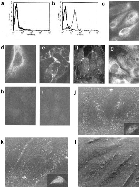
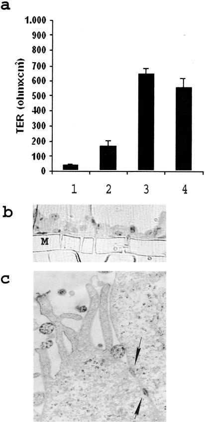
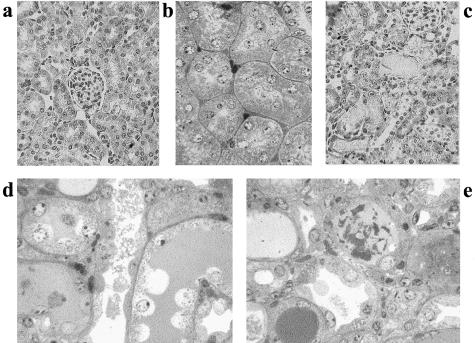
We describe here isolation and characterization of CD133+ cells derived from normal adult human kidney. These cells lacked the expression of hematopoietic markers and expressed PAX-2, an embryonic renal marker, suggesting their renal origin. Renal tissue-derived CD133+ cells and clones of individual cells were capable of expansion and limited self-renewal and differentiated in vitro into epithelial or endothelial cells. On subcutaneous implantation in SCID mice, the undifferentiated cells formed tubular structures expressing renal epithelial markers. At variance, when differentiated in endothelial cells, these cells formed functional vessels. On intravenous injection in SCID mice with glycerol-induced tubulonecrosis, the in vitro expanded renal-derived CD133+ cells homed into the injured kidney and integrated in tubules. We propose that CD133+ cells from kidney represent a multipotent adult resident stem cell population that may contribute to the repair of renal injury.


Figures

Figure 1
Figure 1
Isolation and characterization of CD133+ cells from adult human renal tissue. a and b: Micrographs representative of immunohistochemical detection of CD133+ cells in the cortex of adult normal human kidney. Cells, expressing membrane CD133, are visible within the interstitium (arrows). c and d: Flow cytometric analysis showed that ∼1% of total cells extracted from renal population deprived of glomeruli express CD133. e to i: Flow cytometric analysis of the immunosorted CD133+ cells. Almost all immunosorted cells expressed CD133 (e), CD73 (f), and CD44 (g), but not CD34 (e), CD45 (h), and CD117 (i). Eight different cell preparations were examined with similar results. j: Micrograph representative of Pax-2 nuclear staining in renal progenitor cells. k: PAX-2 mRNA expression by quantitative real-time PCR. Three different cell preparations (▪, •, ♦) were studied in duplicate, CD133+/CD34+ cells from peripheral blood were used as negative control (▴) and cloned PAX-2 as positive control (continuous line). Original magnifications: ×250 (a, j); ×400 (b). G, glomerulus.
Figure 2
Figure 2
Epithelial differentiation of renal tissue-derived CD133+ cells. a and b: Representative cytofluorimetric analysis of CD133+ cells cultured in epithelial differentiating medium. After10 days of culture, cells lost CD133 expression (a), but maintained the expression of CD44 (b). c to i: Representative immunofluorescence micrographs of cytokeratin (c), vimentin (d), E-cadherin (e), ZO-1 (f), alkaline phosphatase (g), and α-smooth muscle actin (h) expression by epithelial differentiated cells. e: Negative control. j to l: Representative micrographs of immunogold detection by scanning electron microscopy and of immunofluorescence (j, inset) of aminopeptidase A (j) and of NaCl co-transporter (k). l: Negative control. Eight cell preparations and 10 clones were studied by immunofluorescence and 2 clones by immunogold scanning electron microscopy with similar results. Original magnifications: ×400 (c-i, insets in j and k); ×1200 (j–l).
Figure 3
Figure 3
Formation of a polarized layer by the epithelial differentiated renal progenitor cells evaluated by the transepithelial resistance and morphological aspects. a: Confluent CD133+ undifferentiated or differentiated cell cultures were plated on Transwell filters and assayed for the transepithelial resistance by an epithelial volt ohm meter. An immortalized tubular epithelial cell line was used as control. Column 1, Semipermeable membrane without cells; column 2, CD133+ undifferentiated cells; column 3, epithelial differentiated renal progenitor cells; column 4, immortalized tubular epithelial cells. Values are expressed as mean ± SD. Ohms/cm2 of three experiments. b: Micrograph representative of toluidine blue-stained semithin section showing a cross section of epithelial differentiated cells cultured in Transwell on a semipermeable membrane (M). c: Transmission electron microscopy micrograph of epithelial differentiated cells cultured in Transwell on a semipermeable membrane showing apical microvilli and junctional complexes (arrows). Original magnifications: ×150 (b); ×20,000 (c).
Figure 4
Figure 4
Endothelial differentiation of renal tissue-derived CD133+ cells. a–f: Representative cytofluorimetric analysis of CD133+ cells cultured in endothelial differentiating medium. After 10 days of culture, cells lost CD133 expression (a); maintained the expression of CD44 (b); and acquired the endothelial markers Muc18 (c), KDR (d), CD105 (e), and VE-cadherin (f). g: The expression of vWF in the classical cytoplasmic punctuate pattern by endothelial differentiated cells, which were negative for cytokeratin (inset) (immunofluorescence micrograph). h–j: Scanning electron microscopy showing spontaneous cell organization on Matrigel. Endothelial differentiated cells (i, j), but not CD133+ undifferentiated cells (h) within 4 hours aligned to form ring-like structures. k: The expression of vWF by differentiated endothelial cells plated on Matrigel. Eight cell preparations and 10 clones were studied with similar results. Original magnifications: ×200 (h, i); × 400 (g, k); ×750 (j).
Figure 5
Figure 5
In vivo differentiation of renal tissue-derived CD133+ cells in tubular structures. Undifferentiated CD133+ cells (2 × 10⁶) at passage III were incorporated at 4°C within Matrigel and subcutaneously injected in SCID mice (n = 6). Mice were sacrificed after 15 days and the Matrigel plugs were excised and processed for histology and immunohistochemistry. a: Micrograph representative of a toluidine blue-stained semithin section showing a transversal section of a tubular structure formed by batiprismatic cells and a central lumen. By immunohistochemistry, the tubules were positive for human HLA class I antigen (inset), for cytokeratin (b), for vimentin (c), for EMA (d), and for NaCl co-transporter (e), but negative for aminopeptidase A (f). g: Some tubules were also positive for alkaline phosphatase. h: By immunofluorescence some tubules showed positive cells for calbindin-D. i: Pax-2 staining was mainly nuclear as observed by immunohistochemistry, in the absence of nuclear counterstaining. Original magnifications: ×150 (b); ×250 (c–i, inset in a and b); ×600 (a).
Figure 6
Figure 6
In vivo differentiation of renal tissue-derived CD133+ cells in tubular structures. Undifferentiated CD133+ cells (2 × 10⁶) at passage III were incorporated at 4°C within Matrigel and subcutaneously injected in SCID mice (n = 6). Mice were sacrificed after 15 days and the Matrigel plugs were excised and processed for electron microscopy. a: Toluidine blue-stained semithin section of a longitudinal section through a tubule showing (right) a lumen partially filled by dense material. On the left tangential section shows cells displaying different staining properties that may depend on different stage of differentiation. b: Low-power electron micrograph showing the arrangement of cells around a virtual lumen (L) filled with dense material. c: Apical part of three adjacent cells showing microvilli (*) and junction complexes (arrows). Original magnifications: ×600 (a); ×6000 (b); ×25,000 (c).
Figure 7
Figure 7
In vivo differentiation of renal tissue-derived CD133+ cells in functional vessels. Endothelial committed cells (2 × 10⁶) at passage III to IV after differentiation were incorporated at 4°C within Matrigel and subcutaneously injected in SCID mice (n = 8). Mice were sacrificed after 15 days and the Matrigel plugs were excised and processed for histology and immunohistochemistry. a: Light microscopy of a trichromic-stained section of Matrigel showing several vessels with patent lumen-containing blood cells. b: The human nature of endothelial cells lining the vessels is indicated by positive immunofluorescence staining for human HLA class I antigen. Nuclei were counterstained in blue with Hoechst 33258 dye. c: Scanning electron microscopy performed on a freeze-hatched Matrigel plug showing a branched vessel containing red cells. Original magnifications: ×400 (a, b); ×1500 (c).
Figure 8
Figure 8
Morphological alterations in glycerol-induced acute tubulonecrosis in SCID mice. a and b: Light micrograph showing normal renal tissue stained with H&E (a) and with toluidine blue. c–e: Tubulonecrotic injury observed 3 days after an intramuscular injection of glycerol (c, H&E-stained section; d and e, toluidine blue-stained semithin sections). Proximal and distal tubules showing loss of brush border, cytoplasmic vacuolization, and flattening of epithelial cells with aspect of apparent denudation of tubular basal membrane. Original magnifications: ×250 (a, c–e); ×400 (b).
Figure 9
Figure 9
In vivo homing of renal tissue-derived CD133+ cells in the kidney of SCID mice with glycerol-induced acute tubulonecrosis. CD133+ cells were injected intravenously in SCID mice 3 days after an intramuscular treatment with glycerol (n = 6) or saline (n = 5). Mice were sacrificed 3 days after cell injection. a–c: Representative micrographs of kidney sections stained with anti-human HLA class I Ab and observed by confocal microscopy. Human HLA class I-positive cells were detected in proximal and distal tubules of mice with glycerol-induced acute tubulonecrosis injected with CD133+ cells (a and c) whereas only minimal positivity was observed in kidney without injury (b). Nuclei were counterstained in blue with Hoechst 33258 dye. d: Proliferating cell nuclear antigen staining of proliferating cells in a field correspondent to part of c. e: Merged image of c and d showing that some of the human HLA class I-positive cells were proliferating cell nuclear antigen-positive (arrows). f–h: Co-staining of cells within a tubule labeled with PHK2 green fluorescent dye before injection (f) and with cytokeratin (g). h: Merge. Original magnifications, ×400. G, glomerulus.

References

    1. Bonventre JV, Weinberg JM. Recent advances in the pathophysiology of ischemic acute renal failure. J Am Soc Nephrol. 2003;14:2199–2210. - PubMed
    1. Kang DH, Kanellis J, Hugo C, Truong L, Anderson S, Kerjaschki D, Schreiner GF, Johnson RJ. Role of the microvascular endothelium in progressive renal disease. J Am Soc Nephrol. 2002;13:806–816. - PubMed
    1. Al-Awqati Q, Oliver JA. Stem cells in the kidney. Kidney Int. 2002;61:387–395. - PubMed
    1. Oliver JA, Barasch J, Yang J, Herzlinger D, Al-Awqati Q. Metanephric mesenchyme contains embryonic renal stem cells. Am J Physiol. 2002;283:F799–F809. - PubMed
    1. Robert B, St. John PL, Abrahamson DR. Direct visualization of renal vascular morphogenesis in Flk1 heterozygous mutant mice. Am J Physiol. 1998;275:F164–F172. - PubMed

Publication types

MeSH terms