Evidence for a role of TNF-related apoptosis-inducing ligand (TRAIL) in the anemia of myelodysplastic syndromes
- PMID: 15681838
- PMCID: PMC1602326
- DOI: 10.1016/S0002-9440(10)62277-8
Evidence for a role of TNF-related apoptosis-inducing ligand (TRAIL) in the anemia of myelodysplastic syndromes
Abstract
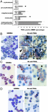
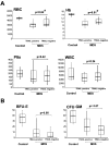
Myelodysplastic syndromes (MDS) are characterized by impaired erythropoiesis, possibly caused by proapoptotic cytokines. We focused our study on the cytokine TRAIL (TNF-related apoptosis-inducing ligand), which has been shown to exhibit an anti-differentiation activity on erythroid maturation. Immunocytochemical analysis of bone marrow mononuclear cells (BMMC) showed an increased expression of TRAIL in MDS patients with respect to acute myeloid leukemia (AML) patients and normal BM donors. TRAIL expression was increased predominantly in myeloid precursors of granulocytic lineage and in a subset of monocytes and pro-erythroblasts. Significant levels of soluble TRAIL were released in 21 of 68 BMMC culture supernatants from MDS patients. On the other hand, TRAIL was detected less frequently in the culture supernatants of AML (4 of 33) and normal BMMC (0 of 22). Analysis of peripheral blood parameters revealed significantly lower levels of peripheral red blood cells and hemoglobin in the subset of patients whose BMMC released TRAIL in culture supernatants compared to the subgroup of patients who did not release TRAIL. Moreover, TRAIL-positive BMMC culture supernatants inhibited the differentiation of normal glycophorin A+ erythroblasts generated in serum-free liquid phase. Thus, increased expression and release of TRAIL at the bone marrow level is likely to impair erythropoiesis and to contribute to the degree of anemia, the major clinical feature of MDS.
Figures


Similar articles
-
The involvement of tumor necrosis factor-related apoptosis-inducing ligand (TRAIL) in atherosclerosis.J Am Coll Cardiol. 2005 Apr 5;45(7):1018-24. doi: 10.1016/j.jacc.2004.12.065. J Am Coll Cardiol. 2005. PMID: 15808757
-
Over-expression of tumor necrosis factor-alpha in bone marrow biopsies from patients with myelodysplastic syndromes: relationship to anemia and prognosis.Eur J Haematol. 2005 Dec;75(6):485-91. doi: 10.1111/j.1600-0609.2005.00551.x. Eur J Haematol. 2005. PMID: 16313260
-
Application of flow cytometry to molecular medicine: detection of tumor necrosis factor-related apoptosis-inducing ligand receptors in acute myeloid leukaemia blasts.Int J Mol Med. 2005 Dec;16(6):1041-8. Int J Mol Med. 2005. PMID: 16273284
-
TNF-related apoptosis-inducing ligand (TRAIL) and erythropoiesis: a role for PKC epsilon.Eur J Histochem. 2006 Jan-Mar;50(1):15-8. Eur J Histochem. 2006. PMID: 16584980 Review.
-
On the TRAIL of a new therapy for leukemia.Leukemia. 2005 Dec;19(12):2195-202. doi: 10.1038/sj.leu.2403946. Leukemia. 2005. PMID: 16224489 Review.
Cited by
-
The sorafenib plus nutlin-3 combination promotes synergistic cytotoxicity in acute myeloid leukemic cells irrespectively of FLT3 and p53 status.Haematologica. 2012 Nov;97(11):1722-30. doi: 10.3324/haematol.2012.062083. Epub 2012 Jun 11. Haematologica. 2012. PMID: 22689683 Free PMC article. Clinical Trial.
-
SOCS1 is significantly up-regulated in Nutlin-3-treated p53wild-type B chronic lymphocytic leukemia (B-CLL) samples and shows an inverse correlation with miR-155.Invest New Drugs. 2012 Dec;30(6):2403-6. doi: 10.1007/s10637-011-9786-2. Epub 2012 Jan 13. Invest New Drugs. 2012. PMID: 22238073
-
Heat Shock Protein 90 is overexpressed in high-risk myelodysplastic syndromes and associated with higher expression and activation of Focal Adhesion Kinase.Oncotarget. 2012 Oct;3(10):1158-68. doi: 10.18632/oncotarget.557. Oncotarget. 2012. PMID: 23047954 Free PMC article.
-
Treating Leukemia in the Time of COVID-19.Acta Haematol. 2021;144(2):132-145. doi: 10.1159/000508199. Epub 2020 May 11. Acta Haematol. 2021. PMID: 32392559 Free PMC article. Review.
-
Targeting of the bone marrow microenvironment improves outcome in a murine model of myelodysplastic syndrome.Blood. 2016 Feb 4;127(5):616-25. doi: 10.1182/blood-2015-06-653113. Epub 2015 Dec 4. Blood. 2016. PMID: 26637787 Free PMC article.
References
-
- Heaney ML, Golde DW. Myelodysplasia. N Engl J Med. 1999;340:1649–1660. - PubMed
-
- Bennett JM. World Health Organization classification of the 399 acute leukemias and myelodysplastic syndrome. Int J Hematol. 2002;72:131–133. - PubMed
-
- Raza A, Gezer S, Mundle S, Gao XZ, Alvi S, Borok R, Rifkin S, Iftikhar A, Shetty V, Parcharidou A. Apoptosis in bone marrow biopsy samples involving stromal and hematopoietic cells in 50 patients with myelodysplastic syndromes. Blood. 1995;86:268–276. - PubMed
-
- Bogdanovic AD, Jankovic GM, Colovic MD, Trpinac DP, Bumbasirevic VZ. Apoptosis in bone marrow of myelodysplastic syndrome patients. Blood. 1996;87:3064–3066. - PubMed
-
- Mundle S, Venugopal P, Shetty V, Ali A, Chopra H, Handa H, Rose S, Mativi BY, Gregory SA, Preisler HD, Raza A. The relative extent and propensity of CD34+ vs. CD34- cells to undergo apoptosis in myelodysplastic marrows. Int J Hematol. 1999;69:152–159. - PubMed
Publication types
MeSH terms
Substances
LinkOut - more resources
Full Text Sources
Medical
Research Materials
Miscellaneous

