. 2005 Feb;166(2):597-613.
doi: 10.1016/s0002-9440(10)62281-x.

Receptor-mediated tobacco toxicity: regulation of gene expression through alpha3beta2 nicotinic receptor in oral epithelial cells

Affiliations

Receptor-mediated tobacco toxicity: regulation of gene expression through alpha3beta2 nicotinic receptor in oral epithelial cells

Juan Arredondo et al. Am J Pathol. 2005 Feb.

Abstract

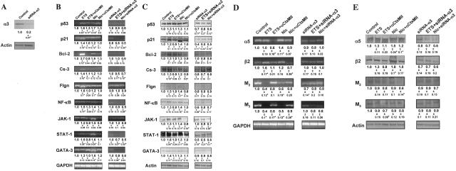
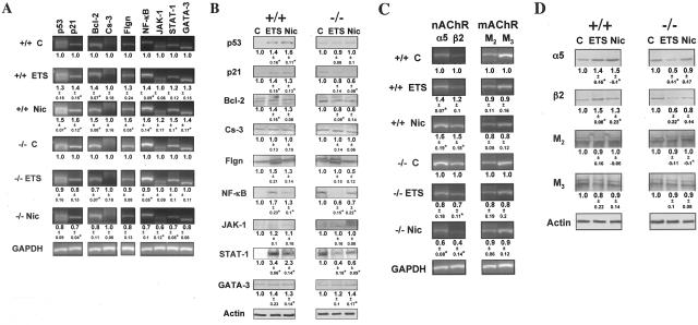
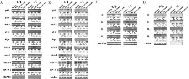
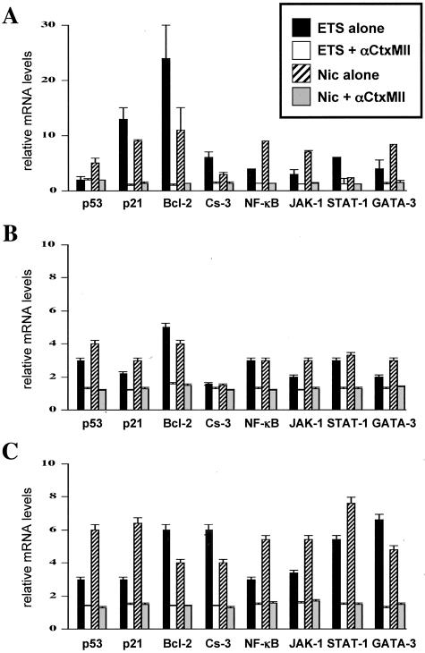
Tobacco is a known cause of oral disease but the mechanism remains elusive. Nicotine (Nic) is a likely culprit of pathobiological effects because it displaces the local cytotransmitter acetylcholine from the nicotinic receptors (nAChRs) expressed by oral keratinocytes (KCs). To gain a mechanistic insight into tobacco-induced morbidity in the oral cavity, we studied effects of exposures to environmental tobacco smoke (ETS) versus equivalent concentration of pure Nic on human and murine KCs. Both ETS and Nic up-regulated expression of cell cycle and apoptosis regulators, differentiation marker filaggrin, and signal transduction factors at both the mRNA and protein levels. These changes could be abolished in cultured human oral KCs transfected with anti-alpha3 small interfering RNA or treated with the alpha3beta2-preferring antagonist alpha-conotoxin MII. Functional inactivation of alpha3-mediated signaling in alpha3-/- mutant KCs prevented most of the ETS/Nic-dependent changes in gene expression. To determine relevance of the in vitro findings to the in vivo situation, we studied gene expression in oral mucosa of neonatal alpha3+/+ and alpha3-/- littermates delivered by heterozygous mice soon after their exposures to ETS or equivalent concentration of pure Nic in drinking water. In addition to reverse transcriptase-polymerase chain reaction and Western blot, the ETS/Nic-dependent alterations in gene expression were also detected by semiquantitative immunofluorescence assay directly in KCs comprising murine oral mucosa. Only wild-type mice consistently developed significant (P < 0.05) changes in the gene expression. These results identified alpha3beta2 nAChR as a major receptor mediating effects of tobacco products on KC gene expression. Real-time polymerase chain reaction demonstrated that in all three model systems the common genes targeted by alpha3beta2-mediated ETS/Nic toxicity were p21, Bcl-2, NF-kappaB, and STAT-1. The expression of the nAChR subunits alpha5 and beta2 and the muscarinic receptor subtypes M(2) and M(3) was also altered. This novel mechanism offers innovative solutions to ameliorate the tobacco-related cell damage and intercede in disease pathways, and may shed light on general mechanisms regulating and driving tobacco-related morbidity in human cells.


Figures

Figure 1
Figure 1
Gene expression changes in human KCs exposed to ETS or pure Nic. Total RNA and proteins were isolated from intact or siRNA-α3-transfected human KCs exposed for 6 hours per day for 5 consecutive days to ETS or 10 μmol/L Nic. The relative amounts of mRNA transcripts and protein levels of the regulatory molecules p53, p21, Bcl-2, caspase-3 (Cs-3), NF-κB, JAK-1, STAT-1, GATA-3, the differentiation markers filaggrin (Flgn), the α5 and β2 nAChR subunits, and the M2 and M3 mAChR subtypes were measured, and the results expressed as described in the Materials and Methods section. Asterisks indicate significant (P < 0.05) differences from control. A: Representative results of WB analysis of the effect of siRNA-α3 on α3 nAChR subunit expression in human KCs. The numbers underneath the bands are ratios of the densitometry value of α3 subunit to that of β-actin, compared to the values obtained in control KCs (taken as 1). B and D: RT-PCR analysis of ETS and Nic effects on gene expression in human KCs. Gene-specific RT-PCR primers were designed to amplify the human p53, p21, Bcl-2, Cs-3, Flgn, NF-κB, JAK-1, STAT-1, and GATA-3 (B), or α3 and β2 nAChR subunit, or M2 and M3 mAChR subtype (D) genes (Table 1). To standardize the analysis, the gene expression ratios in the control cells, ie, intact KCs in experiments with αCtxMII (shown on the gels) and KCs transfected with a nonspecific siRNA in experiments with siRNA-α3 (not shown), were taken as 1. The ratio data underneath the bands are the means ± SD of the values obtained in at least three independent experiments. The images show representative bands in gels. C and E: WB analysis of ETS and Nic effects on gene expression in human KCs. After the exposure experiments described in the B and D, the protein levels of KC p53, p21, Bcl-2, Cs-3, Flgn, NF-κB, JAK-1, STAT-1, and GATA-3 (C), or α3 and β2 nAChR subunits, or M2 and M3 mAChR subtypes (E) were analyzed by WB. The gene expression ratio of 1 was assigned to control KCs, as explained above. The ratio data are the means ± SD of the values obtained in at least three independent experiments. The images show typical bands appearing at the expected molecular weights (Table 4). Specific staining was absent in the negative control experiments in which the membranes were treated without primary antibody or with irrelevant primary antibody of the same isotype and host (not shown).
Figure 2
Figure 2
Null mutation of α3 nAChR subunit abolishes ETS- and Nic-dependent changes in KC gene expression in in vivo experiments. To determine the role of α3 nAChRs in mediating effects of ETS and pure Nic on KCs, the gene expression at the mRNA and protein levels was analyzed using RT-PCR and WB assays, respectively. Asterisks indicate significant (P < 0.05) differences from control. A and C: RT-PCR analysis of ETS and Nic effects on gene expression in oral mucosa of exposed α3−/− mice. Gene-specific RT-PCR primers were designed to amplify the murine p53, p21, Bcl-2, Cs-3, Flgn, NF-κB, JAK-1, STAT-1, and GATA-3 (A), or α3 and β2 nAChR subunit, or M2 and M3 mAChR subtype (C) genes (Table 2). The ratio data underneath the bands are the means ± SD of the values obtained in at least three independent experiments. The images show representative bands in gels. B and D: WB analysis of ETS and Nic effects on gene expression in oral mucosa of exposed α3−/− mice. The proteins levels of p53, p21, Bcl-2, Cs-3, Flgn, NF-κB, JAK-1, STAT-1, and GATA-3 (B), or α3 and β2 nAChR subunits, or M2 and M3 mAChR subtypes (D) were analyzed by WB of total protein isolated from the same α3+/+ and α3−/− mice described in B and D, and analyzed by WB as detailed in the legend to Figure 1, C and E, using primary antibodies listed in Table 4. The gene expression ratio of 1 was assigned to oral tissue samples from α3+/+ mice. The ratio data underneath the bands are the means ± SD of the values obtained in at least three independent experiments. The images show typical bands in gels.
Figure 3
Figure 3
Null mutation of α3 nAChR subunit abolishes ETS- and Nic-dependent changes in KC gene expression in in vitro experiments. Monolayers of murine KCs were established from the oral mucosa of neonatal α3+/+ and α3−/− mice and grown to ∼70% confluence in KGM, after which the monolayers were exposed to ETS or Nic as detailed in Materials and Methods. Asterisks indicate significant (P < 0.05) differences from control. A and C: RT-PCR analysis of ETS and Nic effects on gene expression in exposed α3−/− murine KCs. Gene-specific RT-PCR primers were designed to amplify the murine p53, p21, Bcl-2, Cs-3, Flgn, NF-κB, JAK-1, STAT-1, and GATA-3 (A), or α3 and β2 nAChR subunit, or M2 and M3 mAChR subtype (C) genes (Table 2). To standardize analysis, the gene expression ratio in α3+/+ KCs was taken as 1. The ratio data underneath the bands are the means ± SD of the values obtained in at least three independent experiments. The images show representative bands in gels. B and D: WB analysis of ETS and Nic effects on gene expression in exposed α3−/− murine KCs. The KC proteins under consideration were visualized by primary antibodies (Table 4) using the WB procedure described in detail in the legend to Figure 1, C and E. The gene expression ratio of 1 was assigned to α3+/+ KCs. The ratio data underneath the bands are the means ± SD of the values obtained in at least three independent experiments.
Figure 4
Figure 4
Real-time PCR analysis of the effects of ETS and Nic on KC gene expression revealing the most sensitive targets of the tobacco toxicity. The real-time PCR analysis of the gene expression was performed using RNA isolated from ETS- or Nic-exposed human gingival KCs in the absence of presence of αCtxMII (A), oral mucosa of α3−/−, and α3+/+ littermates (B), and α3−/− and α3+/+ murine oral KCs (C) in experiments described above in the legend to Figures 1, 2, and 3, respectively. The real-time PCR was performed exactly as described in the Materials and Methods section using the primers listed in Table 3. The alterations in the gene expression levels are presented relative to the rates of expression of corresponding genes in control samples, taken as the baseline.

References

    1. Calsina G, Ramon JM, Echeverria JJ. Effects of smoking on periodontal tissues. J Clin Periodontol. 2002;29:771–776. - PubMed
    1. Palmer RM, Scott DA, Meekin TN, Poston RN, Odell EW, Wilson RF. Potential mechanisms of susceptibility to periodontitis in tobacco smokers. J Periodontal Res. 1999;34:363–369. - PubMed
    1. Taybos G. Oral changes associated with tobacco use. Am J Med Sci. 2003;326:179–182. - PubMed
    1. Obeid P, Bercy P. Effects of smoking on periodontal health: a review. Adv Ther. 2000;17:230–237. - PubMed
    1. Chowdhury P, MacLeod S, Udupa KB, Rayford PL. Pathophysiological effects of nicotine on the pancreas: an update. Exp Biol Med (Maywood) 2002;227:445–454. - PubMed

Publication types

MeSH terms