. 2005 Feb;166(2):637-44.
doi: 10.1016/s0002-9440(10)62284-5.

Inhibition of tumor necrosis factor-alpha improves physiological angiogenesis and reduces pathological neovascularization in ischemic retinopathy

Affiliations

Inhibition of tumor necrosis factor-alpha improves physiological angiogenesis and reduces pathological neovascularization in ischemic retinopathy

Tom A Gardiner et al. Am J Pathol. 2005 Feb.

Abstract

The present study was undertaken to test whether inhibition of the proangiogenic inflammatory cytokine tumor necrosis factor (TNF)-alpha can modulate retinal hypoxia and preretinal neovascularization in a murine model of oxygen-induced retinopathy (OIR). OIR was produced in TNF-alpha-/- and wild-type (WT) control C57B6 neonatal mice by exposure to 75% oxygen between postnatal days 7 and 12 (P7 to P12). Half of each WT litter was treated with the cytokine inhibitor semapimod (formerly known as CNI-1493) (5 mg/kg) by daily intraperitoneal injection from the time of reintroduction to room air at P12 until P17. The extent of preretinal neovascularization and intraretinal revascularization was quantified by image analysis of retinal flat-mounts and retinal hypoxia correlated with vascularization by immunofluorescent localization of the hypoxia-sensitive drug pimonidazole (hypoxyprobe, HP). HP adducts were also characterized by Western analysis and quantified by competitive enzyme-linked immunosorbent assay. TNF-alpha-/- and WT mice showed a similar sensitivity to hyperoxia-induced retinal ischemia at P12. At P13 some delay in early reperfusion was evident in TNF-alpha-/- and WT mice treated with semapimod. However, at P17 both these groups had significantly better vascular recovery with less ischemic/hypoxic retina and preretinal neovascularization compared to untreated retinopathy in WT mice. Immunohistochemistry showed deposition of HP in the avascular inner retina but not in areas underlying preretinal neovascularization, indicating that such aberrant vasculature can reduce retinal hypoxia. Inhibition of TNF-alpha significantly improves vascular recovery within ischemic tissue and reduces pathological neovascularization in OIR. HP provides a useful tool for mapping and quantifying tissue hypoxia in experimental ischemic retinopathy.


Figures
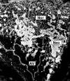
Figure 1
Figure 1
Grading of microvascular configuration in OIR. The retinal microvasculature was localized and identified in retinal flat-mounts using confocal scanning laser microscopy. This representative image shows area of normal intraretinal capillaries (Nor), regions of avascular retina (ischemia, AV), and distinct, hyperfluorescent regions of preretinal neovascularization (Neo).
Figure 2
Figure 2
Semapimod inhibits TNF-α in murine retina. A: Representative Western blot analysis of TNF-α and β-actin in murine retina from nonhyperoxia-exposed mice, OIR mice, and OIR mice treated with semapimod. Data are shown for retinal extracts from P13, P15, and P17 time periods. B: Densitometric analysis of TNF-α Western blot. Data represent the mean ± SD for n = 3 independent experiments. Data are expressed as percentage of control (room air only) mice. *, Significantly different from OIR control (P < 0.05).
Figure 3
Figure 3
OIR-induced retinal ischemia: promotion of intraretinal revascularization by TNF-α inhibition. FITC-dextran angiograms of retinal quadrants from OIR control animals show a complete closure of the central retinal capillary beds (arrows) at P12 (A) (optic disk is indicated by *). B: At P17 semapimod-treated OIR mice show evidence of intraretinal revascularization (RV). C: WT OIR (control) mice at P17 show areas of preretinal neovascularization (arrows), both at the peripheral retina and optic disk (OD). D: In H&E-stained sections there are clear neovascular complexes on the retinal surface (arrow).
Figure 4
Figure 4
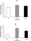
A: TNF-α modulates hyperoxia-induced retinal vascular closure. The areas of avascular retina were quantified in retinal flat-mounts at P13. When WT OIR mice were treated with semapimod there was an increase in the area of avascular retina in comparison to OIR WT controls. This pattern of avascular retina was also evident in TNF-α−/− mice subjected to OIR. Data are the mean ± SD for n = 3 independent experiments (*, P < 0.05). B: Retinal hypoxia is increased with semapimod or absence of TNF-α. Competitive HP ELISA demonstrates increased HP immunoreactivity in retinal lysates from P13 mice for both semapimod-treated mice and TNF-α−/− mice. Data are expressed as the mean concentration per g of total protein (μmol/L/g) ± SD for n = 6 (**, P < 0.01; n = 3 independent experiments).
Figure 5
Figure 5
TNF-α inhibition or deletion reduces retinal neovascularization with concomitant reduction of ischemia and promotion of normal retinal vascularization. Quantitative analysis was performed on the entire retinal microvasculature in flat-mounts at P17. Semapimod treatment and absence of TNF-α resulted in a significant decrease in retinal ischemia (A), an increase in normal (intraretinal) vascularization (B), and a decrease in preretinal neovascularization (C) when compared to WT controls. Data are the mean ± SD for n = 3 independent experiments. **, P < 0.01; ***, P < 0.001.
Figure 6
Figure 6
HP is deposited at sites of retinal hypoxia during OIR. A: At P13 HP deposition is evident (represented by green fluorescence) in the avascular central retina but not in the perfused peripheral retina. Vessels were visualized with isolectin B4 (red fluorescence); optic disk (OD) and neovascularization is apparent at the periphery (NV), but not adjacent to the major retinal arteries (arrow). B: On higher magnification of A (represented by dashed inset) fronds of new vessels (NV) are observed but there is little HP in the underlying retinal neuropile. C: At P17, no HP is evident in the areas underlying neovascularization (NV). D: High magnification of P13 retinal flat-mounts using PI as a nuclear stain reveals high levels of HP immunoreactivity in neurons of the ganglion cell layer (arrows) within nonperfused central retina, remote from traversing arterioles. Original magnifications: ×100 (A, C); ×200 (B, D).

Similar articles

Cited by

References

    1. McCourt M, Wang JH, Sookhai S, Redmond HP. Proinflammatory mediators stimulate neutrophil-directed angiogenesis. Arch Surg. 1999;134:1322–1331. - PubMed
    1. Semenza GL. Regulation of hypoxia-induced angiogenesis: a chaperone escorts VEGF to the dance. J Clin Invest. 2001;108:39–40. - PMC - PubMed
    1. Leibovich SJ, Polverini PJ, Shepard HM, Wiseman DM, Shively V, Nuseir N. Macrophage-induced angiogenesis is mediated by tumour necrosis factor-alpha. Nature. 1987;329:630–632. - PubMed
    1. Frater-Schroder M, Risau W, Hallmann R, Gautschi P, Bohlen P. Tumor necrosis factor type alpha, a potent inhibitor of endothelial cell growth in vitro, is angiogenic in vivo. Proc Natl Acad Sci USA. 1987;84:5277–5281. - PMC - PubMed
    1. BenEzra D, Hemo I, Maftzir G. In vivo angiogenic activity of interleukins. Arch Ophthalmol. 1990;108:573–576. - PubMed

Publication types

MeSH terms