

Inflammatory pseudotumor of the liver and peripheral eosinophilia in autoimmune pancreatitis
- PMID: 15682495
- PMCID: PMC4250611
- DOI: 10.3748/wjg.v11.i6.922
Inflammatory pseudotumor of the liver and peripheral eosinophilia in autoimmune pancreatitis
Abstract
Aim: Inflammatory pseudotumor (IPT) of the liver is a rare benign lesion, the etiology of which remains obscure. It is not associated with any particular diseases apart from phlebitis and Crohn's disease.
Methods: A middle-aged male with hepatic IPT and peripheral eosinophilia associated with autoimmune pancreatitis (AIP) was selected for this study and review of literature.
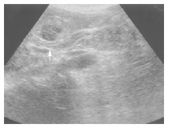
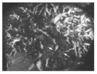
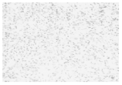
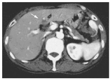
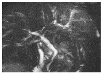
Results: A 59-year-old male was admitted with obstructive jaundice, marked eosinophilia (1 343/mm(3)) and hypergammaglobulinemia (4 145 mg/dL). Imaging techniques revealed dilatation of the intrahepatic bile duct, stenosis of the common bile duct with diffuse wall thickening, gallbladder wall thickening, irregular narrowing of the pancreatic duct, and swelling of the pancreatic parenchyma. Multiple liver masses were also demonstrated and diagnosed as IPT by biopsy specimens. Six months later, the abnormal features of the biliary tree remarkably improved by the oral administration of prednisolone, and the liver masses disappeared. The swelling of the pancreatic head also improved. The peripheral eosinophil count normalized. IPT associated with AIP, as we know, has not been reported in the literature. The clinical features of the present case mimicked those of pancreatic cancer with liver metastasis. This case deserves to be documented to prevent misdiagnosis of similar cases.
Figures

Similar articles
-
Inflammatory pseudotumors of the pancreas and liver with infiltration of IgG4-positive plasma cells.Intern Med. 2007;46(17):1409-12. doi: 10.2169/internalmedicine.46.6430. Epub 2007 Sep 3. Intern Med. 2007. PMID: 17827840
-
Autoimmune pancreatitis, sclerosing cholangitis, liver inflammatory pseudotumors, retroperitoneal fibrosis, lymphadenopathy, and sialoadenitis in a single patient: a multifaceted expression of a hitherto unrecognized systemic autoimmune syndrome?Pancreas. 2009 May;38(4):476-8. doi: 10.1097/MPA.0b013e31818e4793. Pancreas. 2009. PMID: 19390412 No abstract available.
-
Autoimmune pancreatitis with hepatic inflammatory pseudotumor.Pancreas. 2005 Nov;31(4):420-3. doi: 10.1097/01.mpa.0000179732.46210.da. Pancreas. 2005. PMID: 16258381
-
Spontaneous regression of hepatic inflammatory pseudotumor with primary biliary cirrhosis: case report and literature review.World J Gastroenterol. 2006 Mar 14;12(10):1645-8. doi: 10.3748/wjg.v12.i10.1645. World J Gastroenterol. 2006. PMID: 16570364 Free PMC article. Review.
-
A case of concurrent pancreatic intraepithelial neoplasia and type 1 autoimmune pancreatitis with marked pancreatic duct dilatation.Clin J Gastroenterol. 2016 Aug;9(4):266-71. doi: 10.1007/s12328-016-0666-3. Epub 2016 Jun 28. Clin J Gastroenterol. 2016. PMID: 27351197 Review.
Cited by
-
Inflammatory pseudo-tumor of the liver: a rare pathological entity.World J Surg Oncol. 2011 Jan 23;9:5. doi: 10.1186/1477-7819-9-5. World J Surg Oncol. 2011. PMID: 21255461 Free PMC article. Review.
-
Steroid Therapy and Steroid Response in Autoimmune Pancreatitis.Int J Mol Sci. 2019 Dec 30;21(1):257. doi: 10.3390/ijms21010257. Int J Mol Sci. 2019. PMID: 31905944 Free PMC article. Review.
-
Recurrence of inflammatory pseudotumor in the distal bile duct: lessons learned from a single case and reported cases.World J Gastroenterol. 2006 Jun 28;12(24):3938-43. doi: 10.3748/wjg.v12.i24.3938. World J Gastroenterol. 2006. PMID: 16804988 Free PMC article. Review.
-
[Inflammatory myofibroblastic tumor of the lymph node with paraneoplastic thrombosis and eosinophilia].Med Klin (Munich). 2010 Apr;105(4):232-6. doi: 10.1007/s00063-010-1030-x. Med Klin (Munich). 2010. PMID: 20455039 German.
-
Hepatic Inflammatory Pseudotumor-Focusing on Its Heterogeneity.Diagnostics (Basel). 2023 Sep 4;13(17):2857. doi: 10.3390/diagnostics13172857. Diagnostics (Basel). 2023. PMID: 37685395 Free PMC article. Review.
References
-
- Mathiak G, Meyer-Pannwitt U, Mathiak M, Schröder S, Henne-Bruns D, Fröschle G. Inflammatory pseudotumor of the liver--rare differential diagnosis of undetermined hepatic space-occupying lesion. Case report and review of the literature. Langenbecks Arch Chir. 1996;381:309–317. - PubMed
-
- Zamir D, Jarchowsky J, Singer C, Abumoch S, Groisman G, Ammar M, Weiner P. Inflammatory pseudotumor of the liver--a rare entity and a diagnostic challenge. Am J Gastroenterol. 1998;93:1538–1540. - PubMed
-
- Lupovitch A, Chen R, Mishra S. Inflammatory pseudotumor of the liver. Report of the fine needle aspiration cytologic findings in a case initially misdiagnosed as malignant. Acta Cytol. 1989;33:259–262. - PubMed
Publication types
MeSH terms
LinkOut - more resources
Full Text Sources
Medical
Molecular Biology Databases
Miscellaneous
