

Case Reports
doi: 10.3904/kjim.2004.19.4.282.
The first case of primary retroperitoneal mucinous cystadenoma in Korea: a case report
Affiliations
- PMID: 15683120
- PMCID: PMC4531569
- DOI: 10.3904/kjim.2004.19.4.282
Item in Clipboard
Case Reports
The first case of primary retroperitoneal mucinous cystadenoma in Korea: a case report
Korean J Intern Med.
2004 Dec.
Abstract
Primary mucinous cystic cystadenomas of the retroperitoneum are very rarely encountered, and there have been only about 30 cases reported in the literature. The histogenesis of primary mucinous cystadenomas is unclear. Most authors suggested that it develops through mucinous metaplasia in a pre-existing mesothelium-lined cyst. Complete surgical excision is the only treatment and it is required for the final diagnosis and cure. We present here a case report of a 38-year-old Korean woman with primary retroperitoneal cystadenoma. It was a thin-walled, multilocular cyst with a dominant loculus that measured 10.0 x 7.5 x 5.5 cm3 in size, and to the best of our knowledge, this is the first such case to be reported in in Korea.
Figures
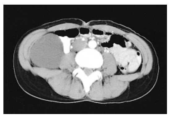
Abdominal CT showed the retroperitoneal mass had displaced the right colon medially and this encapsulated cystic mass contained fluids.
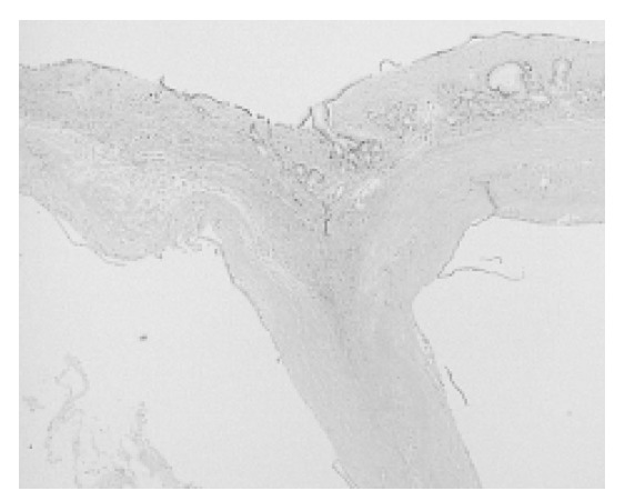
Multilocular cyst lined by a single layer of tall columnar cells. The stroma consisted of fibrocollagenous tissue with no pancreatic or ovarian components (H & E, ×40).
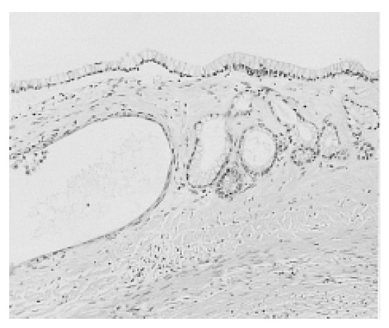
The cells had clear cytoplasm and basely located small nuclei with no cytologic atypia (H & E, ×200).
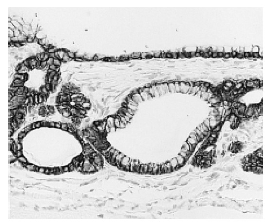
On immunohistochemical staining, the lining cells showed positive reactions to CAM 5.2 (×200).
References
-
- Pennell TC, Gusdon JP., Jr Retroperitoneal mucinous cystadenoma. Am J Obstet Gynecol. 1989;160:1229–1231. - PubMed
-
- Balat O, Aydin A, Sirikci A, Kutlar I, Aksoy F. Huge primary mucinous cystadenoma of the retroperitoneum mimicking a left ovarian tumor. Eur J Gynaecol Oncol. 2001;22:454–455. - PubMed
-
- Yunoki Y, Oshima Y, Murakami I, Takeuchi H, Yasui Y, Tanakaya K, Konaga E. Primary retroperitoneal mucinous cystadenoma. Acta Obstet Gynecol Scand. 1998;77:357–358. - PubMed
-
- Motoyama T, Chida T, Fujiwara T, Watanabe H. Mucinous cystic tumor of the retroperitoneum: a report of two cases. Acta Cytol. 1994;38:261–266. - PubMed
-
- Papadogiannakis N, Gad A, Ehliar B. Primary retroperitoneal mucinous tumor of low malignant potential: histogenetic aspects and review of the literature. APMIS. 1997;105:483–486. - PubMed
Publication types
MeSH terms
LinkOut - more resources
Full Text Sources