. 2005 Jan 31;168(3):415-27.
doi: 10.1083/jcb.200407053.

New GABAergic interneurons in the adult neocortex and striatum are generated from different precursors

Affiliations

New GABAergic interneurons in the adult neocortex and striatum are generated from different precursors

Alexandre G Dayer et al. J Cell Biol. .

Abstract

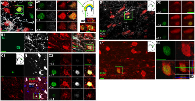
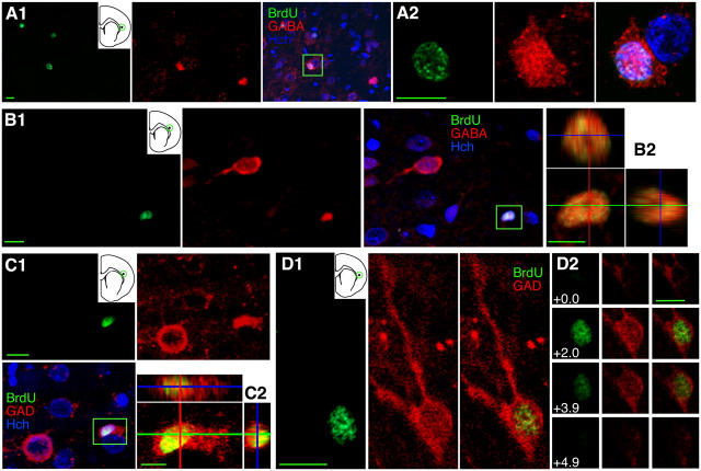
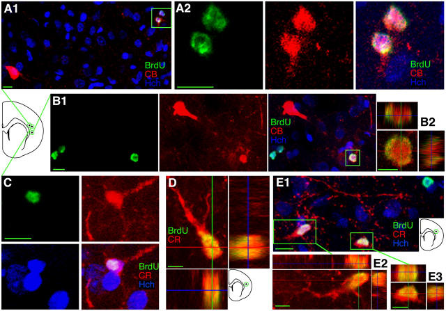
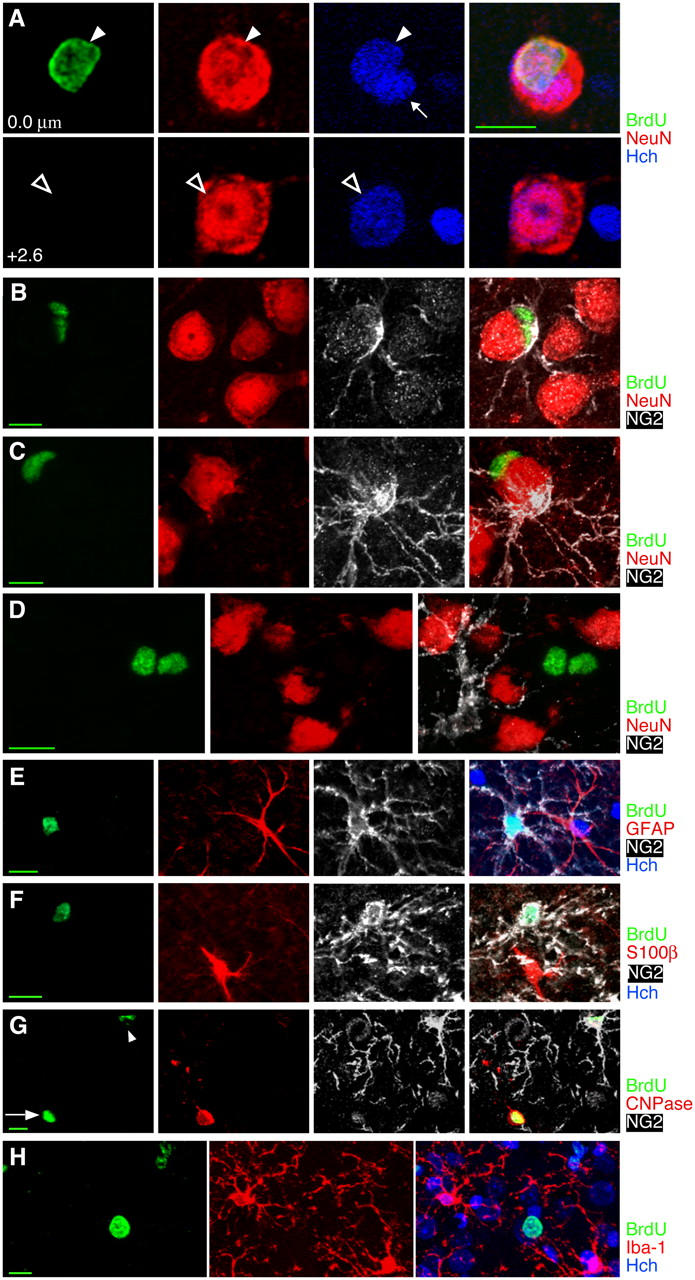
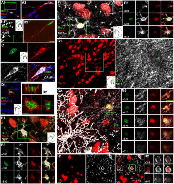
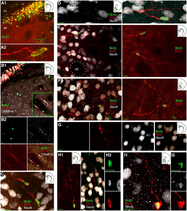
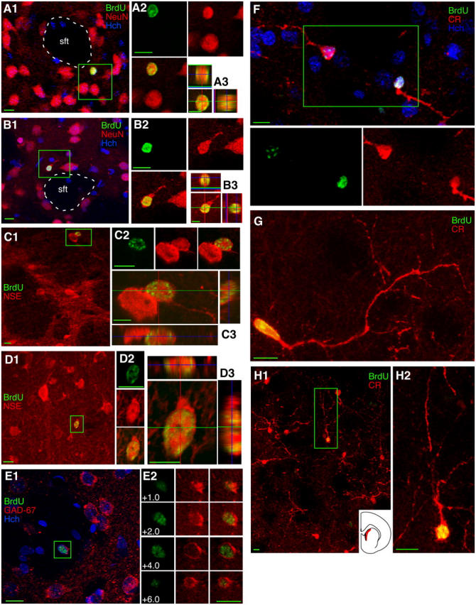
Ongoing neurogenesis in the adult mammalian dentate gyrus and olfactory bulb is generally accepted, but its existence in other adult brain regions is highly controversial. We labeled newly born cells in adult rats with the S-phase marker bromodeoxyuridine (BrdU) and used neuronal markers to characterize new cells at different time points after cell division. In the neocortex and striatum, we found BrdU-labeled cells that expressed each of the eight neuronal markers. Their size as well as staining for gamma-aminobutyric acid (GABA), glutamic acid decarboxylase 67, calretinin and/or calbindin, suggest that new neurons in both regions are GABAergic interneurons. BrdU and doublecortin-immunoreactive (BrdU+/DCX+) cells were seen within the striatum, suggesting migration of immature neurons from the subventricular zone. Surprisingly, no DCX+ cells were found within the neocortex. NG2 immunoreactivity in some new neocortical neurons suggested that they may instead be generated from the NG2+ precursors that reside within the cortex itself.


Figures

Figure 1.
Figure 1.
Some cells born in the adult cortex express neuronal markers. (A) A 4–5-wk-old cell labeled with BrdU and the mature neuronal marker NeuN is shown within the green box in A1. Sample z-planes through the boxed area in A1 are shown with color separation in A2, demonstrating colocalization of BrdU and NeuN but not NG2; note that the green BrdU label and the red NeuN label both peak in brightness at the same level (z = 1.5 μm). The location of the cell shown in A is circled in the small diagram of a coronal section from the appropriate rostrocaudal level (Bregma 1.2); the analyzed region of cortex is shown in yellow on the diagram, and the subcortical white matter, used as a boundary for the analysis, is shown in blue. (B) A 4–5-wk-old BrdU+ (NG2−) neuron labeled with the neuronal marker HuC/D is shown within the box in the multi-channel view in B1 and in orthogonal views in B2. Also note the BrdU+/NG2+/Hu− cell in B1. (C) A 4–5-wk-old BrdU+/NeuN+ neuron that is also immunoreactive for the neuronal glutamate transporter EAAC-1 is seen in color separation in C1, with the blue nuclear counterstain Hoechst 33258 (Hch) added in the multi-color view. Higher magnification views of selected individual z-planes are shown in C2. (D) An 11–12-wk-old BrdU+/NeuN+/NG2− neuron. (E) An 11–12-wk-old BrdU+/NSE+ neuron is shown within the box in E1, at higher magnification with color separation in E2, and in orthogonal views in E3. Images show single focal planes (C1, E1, E2) or Z-axis projections of 14 × 0.64 μm, i.e., 14 planes, 0.64 μm apart (A1), 14 × 0.51 μm (B), 8 × 1.05 μm (D1), or 14 × 0.5 μm (E3). Bars: 5 μm in orthogonal views; 10 μm in all other views.
Figure 2.
Figure 2.
Some cells born in the adult cortex express markers of GABAergic interneurons. (A) A 4–5-wk-old BrdU+/GABA+ cell is shown within the boxed area in the multi-channel view in A1 and at higher magnification in A2. (B) An 11–12-wk-old BrdU+/GABA+ cell in color separation in B1 and in orthogonal views in B2. (C and D) 11–12-wk-old BrdU+/GAD67+ neurons in the cortex. GAD stains cell bodies of some GABAergic neurons but is also strongly expressed in nerve terminals, which appear as GAD+ specks around cell bodies of non-GABAergic neurons. The BrdU+/GABA+ and BrdU/GAD67+ cells in A and C appear to be satellite cells, based on their close apposition to other cells. Images show single focal planes (A2, D1) or Z-axis projections of 4 × 1.71 μm (A1), 5 × 1.17 μm (B), or 7 × 0.87 μm (C). Bars: 5 μm in orthogonal views; 10 μm in all other views.
Figure 3.
Figure 3.
Some cells born in the adult cortex express CB or CR, markers of specific interneuron classes. (A) A pair of 4–5-wk-old BrdU+/CB+ neurons is shown within the box in A1 and at higher magnification color separation in A2. Pairs of BrdU+ labeled cells were frequently observed, suggesting local division from a common precursor. Note the larger BrdU−/CB+ interneuron in the lower left of A1. (B) An 11–12-wk-old BrdU+/CB+ neuron is shown within the box in the multi-channel view in B1 and in orthogonal views in B2. Note the larger BrdU−/CB+ interneuron in the upper left of B1. (C and D) 4–5-wk-old BrdU+/CR+ neurons are shown in color separations in C and (a different cell) in orthogonal views in D. In C, the BrdU+/CR+ neuron and an adjacent BrdU−/CR− cell both appear to be a satellites of a large cell on the left with a speckled nucleus visible in Hoechst staining. (E) A pair of 11–12-wk-old BrdU+/CR+ neurons are shown within the boxes in E1 and in orthogonal views in E2 and E3. Images show Z-axis projections of 6 × 4.96 μm (A1), 10 × 2.03 μm (A2), 7 × 3.17 μm (B1), 9 × 0.84 μm (B2), 14 × 1.47 μm (C), 30 × 0.64 μm (D), 6 × 2.24 μm (E1), 12 × 0.55 μm (E2), or 14 × 0.63 μm (E3). Bars: 5 μm in orthogonal views; 10 μm in all other views.
Figure 4.
Figure 4.
Many cells in the adult rat cortex do not appear to be neurons or commonly recognized glial types. (A) BrdU+ satellite cells closely apposed to large neurons could lead to erroneous identification of the large neurons as BrdU labeled. However, Hoechst nuclear counterstain and z-sectioning make it clear that the 4–5-wk-old BrdU+ cell nucleus (arrowhead at 0.0 μm) is not the nucleus of the large neuron (hollow arrow at 2.6 μm) and must instead belong to a satellite cell. A second, BrdU−, satellite cell can also be seen with Hoechst counterstain (small arrow at 0.0 μm). (B and C) Some but not all 4–5-wk-old BrdU+ satellite cells express the chondroitin sulfate proteoglycan NG2, characteristic of dividing or immature cells. Two satellite cells, one BrdU+/NG2− and one BrdU−/NG2+ can be seen in C. (D) A pair of 4–5-wk-old BrdU+ cells are NG2− and NeuN−, as were ∼40% of 4–5-wk-old BrdU+ cells. (E–G) Newborn NG2+ cells do not stain for astrocyte, oligodendrocyte, or microglial markers. (E) A BrdU+/NG2+ cell and a BrdU−/GFAP+ cell 2 h after BrdU injection. (F) A BrdU+/NG2+ cell and a BrdU−/S100β+ cell 2 h after BrdU injection. (G) A 3–4-wk-old BrdU+ cell in the cortex expressing the oligodendrocyte marker CNPase (arrow) shows faint nuclear staining of NG2 around the nucleus but none of the NG2+ processes that defined NG2+ cells. A second BrdU+ cell (arrowhead) shows strong NG2 staining and very faint CNPase staining. (H) A solidly stained BrdU+ cell and a pair of cells with speckled BrdU staining, all 4–5 wk old and all nonimmunoreactive for the microglial marker Iba-1. Images show single planes (B and E) or Z-axis projections of 10 × 0.69 μm (C), 7 × 1.05 μm (D), 8 × 1.72 μm (F), 9 × 0.67 μm (G), or 18 × 1.38 μm (H). Bars: 5 μm in orthogonal views; 10 μm in all other views.
Figure 5.
Figure 5.
Young cells in the adult cortex expressing CRMP4, NG2, and NeuN, but not DCX, suggest in situ neurogenesis. (A–C) Isolated 1–2-wk-old BrdU+/DCX+ and BrdU+/DCX+/CRMP4+ neurons in the subcortical white matter (wm) dorsal to the striatum (st), have long processes suggesting leading processes. DCX+ cells were relatively easy to find in the SVZ and subcortical white matter but were never seen in the neocortex. (D) A 3–4-wk-old BrdU+/CRMP4+ cell in the cortex shown in the box in D1 and in orthogonal views in D2. (E–G) 4–5-wk-old BrdU+/NeuN+/NG2+ neurons in the cortex suggest a transition from NG2+ cells to NeuN+ cells. NG2 immunoreactivity in processes of BrdU+/NeuN+ cells was either moderate (E and F) or faint (G) and appeared to be inversely correlated with the intensity of NeuN staining. A pair of BrdU+ cells is shown in G2; the strong NG2 staining of the cell on the left nearly obscures its green nucleus. (H) A NeuN+/NG2+ neuron in a section not immunostained for BrdU indicates that this coexpression is not related to BrdU labeling. Images show single a focal plane (H) or Z-axis projections of 7 × 0.92 μm (A1), 14 × 0.58 μm (A2), 10 × 2.28 μm (B1), 15 × 1.5 μm (B2), 8 × 0.96 μm (C), 9 × 1.08 μm (D1), 16 × 0.57 μm (D2), 12 × 0.45 μm (E1), 9 × 0.78 μm (F1), 16 × 3.35 μm (G1), or 16 × 0.47 μm (G2). Bars: 5 μm in orthogonal views; 10 μm in all other views.
Figure 6.
Figure 6.
Young neurons appear to migrate from the adult SVZ into the striatum. (A and B) A band of DCX+ and CRMP4+ 1–2-wk-old BrdU+ neurons appears in the extension of the SVZ between the striatum (str) and the corpus callosum (cc). Scattered immature neurons can be seen outside of this band, either within the striatal matrix as the cell boxed in A1 or in a striatal fiber tract as one of the boxed cells in B1. (C) 1–2-wk-old BrdU+/DCX+ neurons in a striatal fiber tract. (D) A 1–2-wk-old BrdU+/DCX+ neuron in the core of the nucleus accumbens. (E and F) 3–4-wk-old BrdU+/DCX+ neurons extending complex processes are seen in the striatal matrix outside striatal fiber tracts (sft) as in E and in the shell of the nucleus accumbens as in F. (G–I) 3–4-wk-old BrdU+/DCX+/NeuN+ neurons can be found in the striatum as in G and H as well as in the shell of the nucleus accumbens as in I. Images show Z-axis projections of 12 × 0.90 μm (A1); 16 × 0.85 μm (A2); 18 × 0.72 μm (B1), 30 × 0.91 μm (B2), 18 × 1.44 μm (C), 16 × 0.78 μm (D), 36 × 0.48 μm (E), 20 × 0.98 μm (F); 14 × 1.04 μm (G); 30 × 0.78 μm (H1), 11 × 0.78 μm (H2), 16 × 0.62 μm (I1 and I2). Bars: 5 μm in orthogonal views; 10 μm in all other views.
Figure 7.
Figure 7.
BrdU-labeled cells in the adult striatum express markers of GABAergic interneurons. (A) A 4–5-wk-old BrdU+ cell expressing the neuronal marker NeuN in the striatum just outside a striatal fiber tract (sft) is seen in the box in A1, in color separations in A2, and in orthogonal views in A3. (B) An 11–12-wk-old striatal BrdU+/NeuN+ neuron is shown in the same three formats as the cell in A. (C and D) 11–12-wk-old striatal BrdU+/NSE+ neurons were seen near the border of the white matter, as the boxed cell in C1, or deeper within the striatum, as the boxed cell in D1. (E) An 11–12-wk-old BrdU+/GAD67+ neuron in the striatum is shown with surrounding neurons in E1 and in color and z-plane separations in E2. (F–H) 4–5-wk-old BrdU+/CR+ neurons were found in the striatum, and nearly all newborn striatal neurons were found in a region known to have a high density of CR+ neurons, seen in red on the section outline (H1, inset). Images show single focal planes (A1, A2, B2, C1, C2, D2) or Z-axis projections of 10 × 1.02 μm (A3), 10 × 0.92 μm (B1, B3), 10 × 0.65 μm (C3), 6 × 0.71 μm (D1, D3), 7 × 1.20 μm (E1), 18 × 0.74 μm (F), 20 × 0.63 μm (G), 14 × 2.63 μm (H1), 18 × 0.69 μm (H2). Bars: 5 μm in orthogonal views; 10 μm in all other views.

References

    1. Aguirre, A.A., R. Chittajallu, S. Belachew, and V. Gallo. 2004. NG2-expressing cells in the subventricular zone are type C-like cells and contribute to interneuron generation in the postnatal hippocampus. J. Cell Biol. 165:575–589. - PMC - PubMed
    1. Alonso, G. 2000. Prolonged corticosterone treatment of adult rats inhibits the proliferation of oligodendrocyte progenitors present throughout white and gray matter regions of the brain. Glia. 31:219–231. - PubMed
    1. Altman, J. 1962. Are new neurons formed in the brains of adult mammals? Science. 135:1127–1128. - PubMed
    1. Bedard, A., M. Cossette, M. Levesque, and A. Parent. 2002. Proliferating cells can differentiate into neurons in the striatum of normal adult monkey. Neurosci. Lett. 328:213–216. - PubMed
    1. Belachew, S., R. Chittajallu, A.A. Aguirre, X. Yuan, M. Kirby, S. Anderson, and V. Gallo. 2003. Postnatal NG2 proteoglycan-expressing progenitor cells are intrinsically multipotent and generate functional neurons. J. Cell Biol. 161:169–186. - PMC - PubMed

Publication types

MeSH terms

Substances