. 2005 Jan 31;168(3):441-52.
doi: 10.1083/jcb.200407076.

Molecular mechanisms of invadopodium formation: the role of the N-WASP-Arp2/3 complex pathway and cofilin

Affiliations

Molecular mechanisms of invadopodium formation: the role of the N-WASP-Arp2/3 complex pathway and cofilin

Hideki Yamaguchi et al. J Cell Biol. .

Abstract

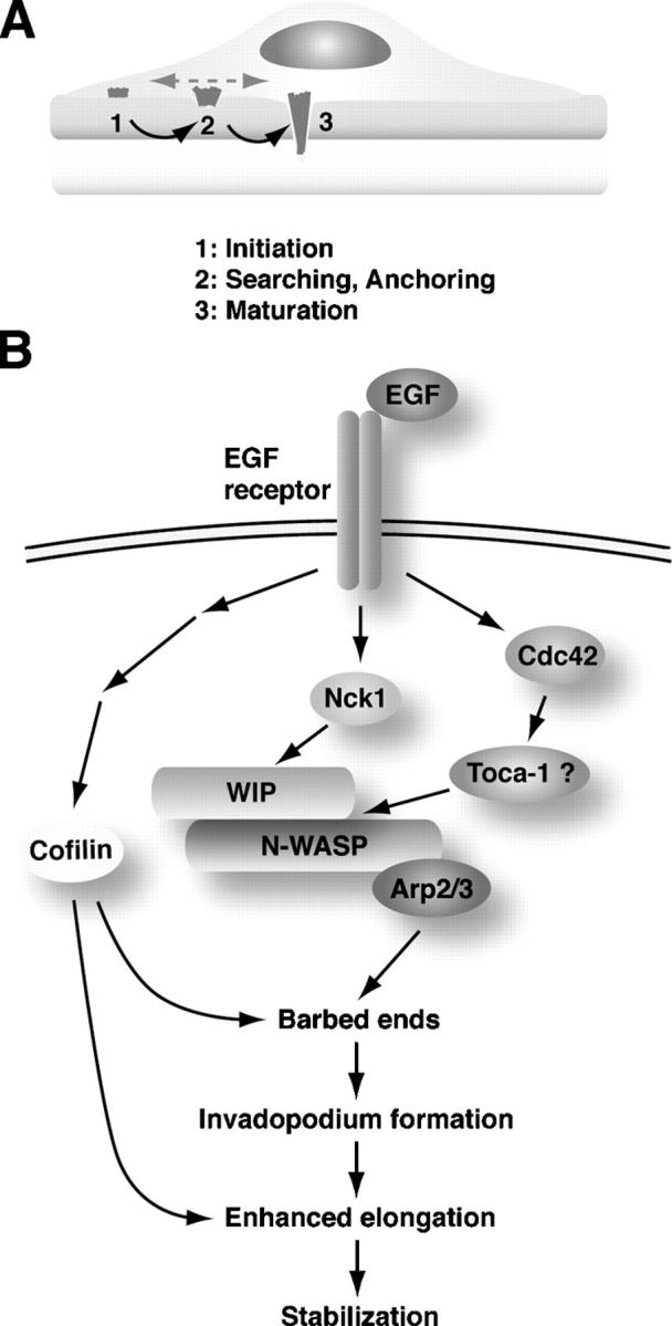
Invadopodia are actin-rich membrane protrusions with a matrix degradation activity formed by invasive cancer cells. We have studied the molecular mechanisms of invadopodium formation in metastatic carcinoma cells. Epidermal growth factor (EGF) receptor kinase inhibitors blocked invadopodium formation in the presence of serum, and EGF stimulation of serum-starved cells induced invadopodium formation. RNA interference and dominant-negative mutant expression analyses revealed that neural WASP (N-WASP), Arp2/3 complex, and their upstream regulators, Nck1, Cdc42, and WIP, are necessary for invadopodium formation. Time-lapse analysis revealed that invadopodia are formed de novo at the cell periphery and their lifetime varies from minutes to several hours. Invadopodia with short lifetimes are motile, whereas long-lived invadopodia tend to be stationary. Interestingly, suppression of cofilin expression by RNA interference inhibited the formation of long-lived invadopodia, resulting in formation of only short-lived invadopodia with less matrix degradation activity. These results indicate that EGF receptor signaling regulates invadopodium formation through the N-WASP-Arp2/3 pathway and cofilin is necessary for the stabilization and maturation of invadopodia.


Figures

Figure 1.
Figure 1.
Metastatic MTLn3 cells but not nonmetastatic MTC cells form invadopodia. (A) MTLn3 cells cultured on Alexa488-FN– and gelatin-coated glass coverslips were stained with rhodamine phalloidin. Bottom images are XZ sections showing a cell with an invadopodium collected with a confocal microscope. Bar, 10 μm. (B) MTC and MTLn3 cells grown on FN- and gelatin-coated coverslips were stained with anti-cortactin antibody and phalloidin. Arrowheads indicate cells displaying invadopodia. Bar, 20 μm. (C) MTC cells cultured on Alexa488-FN (green)– and gelatin-coated glass coverslips were stained with rhodamine phalloidin (red). Bar, 10 μm. (D) The percentage of cells with invadopodia in MTC and MTLn3 cells was calculated as described in Materials and methods. Error bars represent the SD of three different determinations.
Figure 2.
Figure 2.
Time-lapse analysis of invadopodium formation. (A) Time-lapse image sequence of MTLn3 cells expressing GFP-actin. Cells were cultured on FN-gelatin–coated coverslips and analyzed by automated time-lapse microscopy. The invadopodium denoted by an arrowhead is stationary, whereas invadopodia in the lower right cell are dynamic. Bar, 10 μm. (a–c) Time-lapse image sequences of boxed regions. (B) Time-lapse image sequence showing a long-lived stationary invadopodium (closed arrowhead). Open arrowhead denotes an invadopodium left behind by a moving cell. Bar, 10 μm. (C) The fluorescence intensity of the invadopodium shown in Fig. 2 A (a) was plotted versus the speed of its movement as a function of time. The inverse correlation between the fluorescence intensity and the speed indicates that actin polymerization occurs in invadopodia when they become stationary. (D) Distribution of the lifetimes of invadopodia shown in histogram. (E and F) The trajectories of representative long-lived (E) and short-lived (F) invadopodia are shown as lines. The lifetime of each invadopodium is also shown in the box. Bar, 5 μm. (G) Net distance moved by each invadopodium was calculated as shown in the inset and divided by its lifetime, and the ratio was plotted versus lifetime. The ratio is expected to be lower if invadopodia are more stationary. The data indicate that long-lived invadopodia are more stationary than short-lived invadopodia.
Figure 3.
Figure 3.
EGF and EGF receptor signaling are necessary for invadopodium formation. (A) Effect of an EGF receptor kinase inhibitor AG1478 on invadopodium formation. Cells were cultured on FN-gelatin coverslips in the presence of serum with or without AG1478 for 16 h. Cells were then stained with rhodamine phalloidin and anti-cortactin antibody to quantitate invadopodium formation. (B) Effect of serum starvation and subsequent EGF stimulation on invadopodium formation. Cells cultured on FN-gelatin coverslips were serum starved for 4 h with or without 5 μM AG1478 and stimulated with 12.5 nM EGF for 4 min. Error bars represent the SD of three different determinations. (C) Morphology of cells treated with AG1478. After treatment with AG1478 in the presence of serum, cells were stained with rhodamine phalloidin and anti-cortactin antibody. Cells formed stress fibers and lamellipodia even after AG1478 treatment. (D) Morphology of cells serum starved and stimulated with EGF. Arrowheads denote invadopodia. Bars, 10 μm.
Figure 4.
Figure 4.
Localization of WASP family proteins and Arp2/3 complex at invadopodia. Cells grown on FN-gelatin–coated coverslips were stained with anti-N-WASP, anti-WAVE1, anti-WAVE2, or anti-p34arc antibody. To visualize actin filaments, the cells were also stained with rhodamine phalloidin. Arrowheads denote invadopodia. Bar, 10 μm.
Figure 5.
Figure 5.
N-WASP and Arp2/3 complex are involved in invadopodium formation. (A) Cells were transfected with control, N-WASP, WAVE1, WAVE2, or p34arc siRNA. Cell lysates were prepared at 48 h after transfection and immunoblotted with the indicated antibodies. Anti-actin antibody was used for loading controls. (B) Cells treated with siRNAs were stained with anti-cortactin and rhodamine phalloidin to visualize invadopodia (arrowheads). Bar, 10 μm. (C) Invadopodium formation in cells treated with siRNAs. For rescue experiments, N-WASP siRNA-treated cells were transfected with the bovine GFP-N-WASP construct. (D) Invadopodium formation in cells infected with retroviruses expressing control GFP or dominant-negative N-WASP (NW Δcof). (E) Cells were transfected with control GFP or GFP-N-WASP CA constructs, and invadopodium formation was quantified. Error bars represent the SD of three different determinations.
Figure 6.
Figure 6.
Nck1 but not Grb2 is required for formation of invadopodia. (A) Cells transfected with GFP-Nck1 or Grb2 construct were stained with rhodamine phalloidin to visualize invadopodia (arrowheads). Bar, 10 μm. (B) Cells were transfected with control, Nck1, or Grb2 siRNA. Cell lysates were immunoblotted with anti-Nck, anti-Grb2, and anti-actin antibodies. (C) Invadopodia formation was quantified among cells treated with indicated siRNAs. Nck1 siRNA-treated cells were transfected with human GFP-Nck1 construct for rescue experiment. Error bars represent the SD of three different determinations.
Figure 7.
Figure 7.
Cdc42 and WIP–N-WASP interaction are essential for invadopodium formation. (A) Cells were transfected with control or Cdc42 siRNA, and the cell lysates were immunoblotted with anti-Cdc42 and anti-actin antibodies. Cdc42 was not detected in cells treated with Cdc42 siRNA. (B) Cells treated with control or Cdc42 siRNA were stained with anti-cortactin and rhodamine phalloidin to visualize invadopodia. (C) Invadopodia formation was quantified among cells transfected with control or Cdc42 siRNA. (D) Cells transfected with GFP-tagged WIP wild type (WT), N-WASP binding domain (WBD), or cortactin binding domain (CBD) construct were stained with rhodamine phalloidin. (E) Invadopodia formation was quantified among transfected cells. (F) Cells expressing GFP-tagged wild-type WISH (WT) or WISH mutant lacking SH3 domain (ΔSH3) were stained with rhodamine phalloidin. (G) Quantitation of invadopodium formation. Error bars represent the SD of three different determinations. Arrowheads denote invadopodia. Bars, 10 μm.
Figure 8.
Figure 8.
Cofilin is required for fully functional invadopodia. (A) Cells grown on FN-gelatin–coated coverslips were stained with anti-cofilin antibody and rhodamine phalloidin. Arrowheads denote invadopodia. (B) Cells were transfected with control or cofilin siRNA, and the cell lysates were immunoblotted with anti-cofilin and anti-actin antibodies. (C) Cells treated with control or cofilin siRNA were stained with anti-cortactin antibody and rhodamine phalloidin. Arrowheads denote invadopodia. (D) Invadopodia formation was quantified among cells transfected with control or cofilin siRNA. Error bars represent the SD of three different determinations. Asterisk denotes statistical significance (P < 0.04) compared with control calculated by t test. (E) Cells treated with control or cofilin siRNA were cultured on Alexa488-FN– and gelatin-coated glass coverslips to analyze their degradation activity. The cells were stained with rhodamine phalloidin. Arrowheads denote the degradation sites. (F) MTLn3 cells expressing YFP-actin were transfected with control or cofilin siRNA and analyzed by time-lapse microscopy. Lifetime of invadopodia was calculated from the time-lapse movies and shown in histograms. Bars, 10 μm.
Figure 9.
Figure 9.
Model for invadopodium formation. (A) A model for invadopodium formation. Mature invadopodia are formed through three processes. (1) In the initiation phase, precursors of invadopodia are formed de novo at the cell periphery around lamellipodia. (2) These precursors are motile and move around on the ventral membrane until they stick to ECM. (3) In the maturation phase, anchored invadopodia protrude into ECM and degrade it. (B) Schematic diagram of the signaling pathway for invadopodium formation. EGF ligand binding to the EGF receptor activates and/or recruits Nck1 and Cdc42. Nck1 recruits WIP–N-WASP complex, and Cdc42 activates N-WASP probably through Toca-1 to initiate invadopodium formation. Cofilin mainly functions in the maturation process of invadopodium formation.

References

    1. Bailly, M., L. Yan, G.M. Whitesides, J.S. Condeelis, and J.E. Segall. 1998. Regulation of protrusion shape and adhesion to the substratum during chemotactic responses of mammalian carcinoma cells. Exp. Cell Res. 241:285–299. - PubMed
    1. Baldassarre, M., A. Pompeo, G. Beznoussenko, C. Castaldi, S. Cortellino, M.A. McNiven, A. Luini, and R. Buccione. 2003. Dynamin participates in focal extracellular matrix degradation by invasive cells. Mol. Biol. Cell. 14:1074–1084. - PMC - PubMed
    1. Banzai, Y., H. Miki, H. Yamaguchi, and T. Takenawa. 2000. Essential role of neural Wiskott-Aldrich syndrome protein in neurite extension in PC12 cells and rat hippocampal primary culture cells. J. Biol. Chem. 275:11987–11992. - PubMed
    1. Benesch, S., S. Lommel, A. Steffen, T.E. Stradal, N. Scaplehorn, M. Way, J. Wehland, and K. Rottner. 2002. Phosphatidylinositol 4,5-biphosphate (PIP2)-induced vesicle movement depends on N-WASP and involves Nck, WIP, and Grb2. J. Biol. Chem. 277:37771–37776. - PubMed
    1. Buccione, R., J.D. Orth, and M.A. McNiven. 2004. Foot and mouth: podosomes, invadopodia and circular dorsal ruffles. Nat. Rev. Mol. Cell Biol. 5:647–657. - PubMed

Publication types

MeSH terms

Substances