. 2005 Feb 8;102(6):2180-5.
doi: 10.1073/pnas.0409803102. Epub 2005 Jan 31.

The fragile X mental retardation protein and group I metabotropic glutamate receptors regulate levels of mRNA granules in brain

Affiliations

The fragile X mental retardation protein and group I metabotropic glutamate receptors regulate levels of mRNA granules in brain

Armaz Aschrafi et al. Proc Natl Acad Sci U S A. .

Abstract

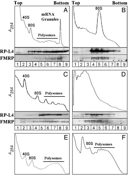
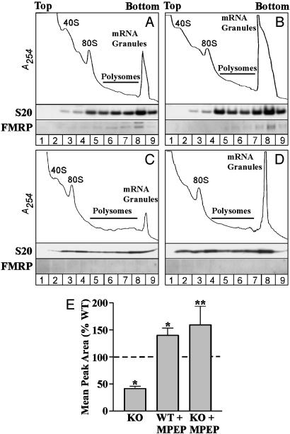
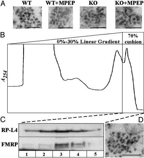
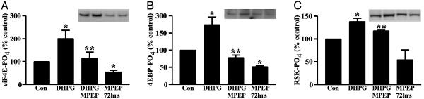
Fragile X syndrome results from the transcriptional silencing of a gene, Fmr1, that codes for an mRNA-binding protein (fragile X mental retardation protein, FMRP) present in neuronal dendrites. FMRP can act as a translational suppressor, and its own translation in dendrites is regulated by group I metabotropic glutamate receptors (mGluRs). Multiple lines of evidence suggest that mGluR-induced translation is exaggerated in Fragile X syndrome because of a lack of translational inhibition normally provided by FMRP. We characterized the role of FMRP in the regulation of mRNA granules, which sediment as a heavy peak after polysomes on sucrose gradients. In WT mouse brain, FMRP distributed with polysomes and granules. EM and biochemical analyses suggested that the granule fraction itself contained clusters of polysomes. In Fmr1 knockout brain, we observed a significant decrease in the amount of mRNA granules relative to WT mice. This difference appeared to be due to a role of FMRP in regulating the activation of granules during mGluR-induced translation; in vivo administration of the mGluR5 antagonist 2-methyl-6-(phenylethynyl)pyridine increased granule content in Fmr1 knockout mouse brain to levels comparable with those seen in WT brain. In accord with a role of mGluR5 in the regulation of ongoing translation in vivo, we observed that the phosphorylation of several initiation factors in response to application of the mGluR1/5 agonist S-3,5-dihydroxyphenylglycine in vitro was blocked by methyl-6-(phenylethynyl)pyridine. Together, these data suggest that although large, polysome-containing granules can form in the absence of FMRP, their use in response to mGluR-induced translation is exaggerated.


Figures

Fig. 1.
Fig. 1.
FMRP cofractionates with polysomes and heavy mRNA granules in lysates of postnatal day 13 rat brain. (A) Absorbance profile (254 nm) of rat brain postmitochondrial fraction sedimented through a 20 -55% linear sucrose gradient in the presence of cyclohexamide. Western blots of samples taken throughout the gradient (numbered at the bottom) show that ribosomal protein L4 (RP-L4) and FMRP are in the polysomes and a heavier granule fraction. The heavy mRNA granule fraction was eliminated by treatment of the lysates with RNase A (B), heating at 37°C (C), or treatment with 30 mM EDTA (D) before fractionation. Granules were not observed in lysates of liver (E) or testis (F).
Fig. 2.
Fig. 2.
mRNA granules are reduced in Fmr1 KO mouse brain, relative to WT, and they are reconstituted by antagonism of mGluR5 in vivo. Absorbance profiles of mRNA granules resolved from WT and Fmr1 KO mice show that, relative to WT animals (A), Fmr1 KO mice (C) have a reduced mRNA granule peak. Injection of the mGluR5-specific antagonist MPEP before killing increased the mRNA granule peak in WT (B) and Fmr1 KO (D) mice. Results varied across experiments, but consistent reductions in the KO and increases with MPEP were observed in each experiment. (E) Bar graph showing the average granule peak area, expressed as a percentage of the WT, in Fmr1 KO mice and in both genotypes after injection of MPEP. Significant differences were observed in comparisons of Fmr1 KO (P < 0.01, n = 12) and MPEP-injected WT mice (P < 0.05, n = 6) versus WT (n = 17) and of Fmr1 KO mice with and without MPEP (P < 0.01, n = 5) (*, one-sample t test; **, two-tailed t test).
Fig. 3.
Fig. 3.
EM examination of granules resolved from WT and Fmr1 KO mouse brain. (A) Electron micrographs of granules collected from WT, KO, and MPEP-injected animals. (Scale bar, 100 nm.) mRNA granules are shown as large, densely packed clusters of 10 -20 ribosomes, ranging in size from 100 to 300 nm. (B) Velocity-gradient isolation of granules from brains lysed without detergent according to the method of Kanai et al. (28). Large particles that absorb at 254 nm penetrate a dense 70% sucrose cushion after separation from other translation components on a 15-30% gradient. (C) Western blot analysis of fractions from the 70% cushion show that FMRP cosediments with these large particles and the ribosomal protein L4. (D) EM analysis revealed large, densely packed clusters of ribosomes that ranged in size from 100 to 300 nm, resembling the granules resolved on 20 -55% linear gradients in the presence of detergent, as shown in A.
Fig. 4.
Fig. 4.
Modulation of translation factor phosphorylation by the mGluR5 antagonist, MPEP. Hippocampal slice cultures were treated with DHPG for 7.5 min with or without MPEP, or with MPEP alone for 72 h. DHPG increased the phosphorylation of eIF4E (A), eIF4E-BP (B), and the 90-kDa ribosome-specific kinase, p90RSK (C). (Insets) Blots show examples of changes in the phosphorylation of each protein. Their order follows the graph conditions. Phosphorylation levels in DHPG-treated slices were significantly different from untreated slices (P < 0.05) or those coincubated with MPEP (P < 0.05). MPEP alone significantly decreased phosphorylation of eIF4E and eIF4E-BP relative to controls. P < 0.05, for one-sample (*) and one-tailed (**) t test. All data reflect averages from four to seven independent experiments with internal replicates.

References

    1. O'Donnell, W. T. & Warren, S. T. (2002) Annu. Rev. Neurosci. 25, 315-338. - PubMed
    1. Hagerman, R. J., Amiri, K. & Cronister, A. (1991) Am. J. Med. Genet. 38, 283-287. - PubMed
    1. Hinton, V. J., Brown, W. T., Wisniewski, K. & Rudelli, R. D. (1991) Am. J. Med. Genet. 41, 289-294. - PubMed
    1. Irwin, S. A., Galvez, R. & Greenough, W. T. (2000) Cereb. Cortex 10, 1038-1044. - PubMed
    1. Comery, T. A., Harris, J. B., Willems, P. J., Oostra, B. A., Irwin, S. A., Weiler, I. J. & Greenough, W. T. (1997) Proc. Natl. Acad. Sci. USA 94, 5401-5404. - PMC - PubMed

Publication types

MeSH terms

Substances

LinkOut - more resources