. 2005 Feb 7;201(3):349-60.
doi: 10.1084/jem.20041542. Epub 2005 Jan 31.

CD8+ immunodominance among Epstein-Barr virus lytic cycle antigens directly reflects the efficiency of antigen presentation in lytically infected cells

Affiliations

CD8+ immunodominance among Epstein-Barr virus lytic cycle antigens directly reflects the efficiency of antigen presentation in lytically infected cells

Victoria A Pudney et al. J Exp Med. .

Abstract

Antigen immunodominance is an unexplained feature of CD8+ T cell responses to herpesviruses, which are agents whose lytic replication involves the sequential expression of immediate early (IE), early (E), and late (L) proteins. Here, we analyze the primary CD8 response to Epstein-Barr virus (EBV) infection for reactivity to 2 IE proteins, 11 representative E proteins, and 10 representative L proteins, across a range of HLA backgrounds. Responses were consistently skewed toward epitopes in IE and a subset of E proteins, with only occasional responses to novel epitopes in L proteins. CD8+ T cell clones to representative IE, E, and L epitopes were assayed against EBV-transformed lymphoblastoid cell lines (LCLs) containing lytically infected cells. This showed direct recognition of lytically infected cells by all three sets of effectors but at markedly different levels, in the order IE > E >> L, indicating that the efficiency of epitope presentation falls dramatically with progress of the lytic cycle. Thus, EBV lytic cycle antigens display a hierarchy of immunodominance that directly reflects the efficiency of their presentation in lytically infected cells; the CD8+ T cell response thereby focuses on targets whose recognition leads to maximal biologic effect.


Figures

Figure 1.
Figure 1.
Assays on EBV-specific CD8 + T cell clones from IM 73. (A) Representative antigen mapping results from 1 of 15 BRLF1-specific clones, 1 of 4 BMLF1-specific clones, and 1 of 7 BALF2-specific clones tested on autologous LCL targets preinfected with the panel of 23 recombinant vaccinias expressing individual IE, E, and L proteins of the EBV lytic cycle. (B) Epitope mapping results from the 26 EBV lytic antigen-specific clones tested on autologous LCL targets preexposed to the BRLF1-derived YVL and IACP peptides, to the BMLF1-derived GLC peptide, and to the BALF2-derived ARYA peptide (restriction elements as shown). All results are expressed as a percentage of specific lysis in 5-h chromium release assays.
Figure 2.
Figure 2.
Assays on EBV-specific CD8 + T cell clones from IM 179. (A) Representative antigen mapping results from 1 of 12 BRLF1-specific clones, 1 of 4 BMRF1-specific clones, 1 BMLF1-specific clone, 1 of 4 BALF2-specific clones, 1 of 4 BALF4-specific clones, and 1 BILF2-specific clone. (B) Epitope mapping results from the 26 EBV lytic antigen–specific clones tested on autologous LCL targets preexposed to the BRLF1-derived YVL and IACP peptides, to the BMRF1-derived TLD peptide, to the BMLF1-derived GLC peptide, to the BALF2-derived ARYA peptide, to the BALF4- derived FLD peptide, and to the BILF2-derived RRRK peptide (restriction elements as shown). Results expressed as in Fig. 1.
Figure 3.
Figure 3.
Summary of antigen mapping data from 11 acute IM patients (HLA class I types shown). A mean of 20 EBV-specific clones were analyzed per patient. For each patient, the EBV lytic antigens recognized by CD8+ T cell clones are identified as shaded boxes, and the percentage of CD8+ T cell clones from each donor recognizing each target antigen is reflected by the intensity of the shading. Note that in two cases, IM 179 and IM 140, clonings were also performed without γ-irradiated autologous LCL feeders, either using a γ-irradiated allogeneic BZLF1-LCL feeder that could not provide any lytic antigen–specific stimulus or using an anti-CD3 mAb as a pan-T cell stimulus. Both protocols yielded the same spectrum of antigen specificities in proportions that were not significantly different from those illustrated above. Combining the results from these alternative protocols, for IM 179 (of 20 clones) 50% clones were IE antigen specific, 25% clones E antigen specific, and 25% clones L antigen specific; for IM 140.1, (of 30 clones) 90% clones were IE antigen specific and 10% E antigen specific.
Figure 4.
Figure 4.
Antigen presentation assays using two CD8 + T cell clones specific for the BZLF1-derived RAK peptide (HLA-B*0801 restricted). A and B show data from an ELISPOT assay on the BZLF1+ LCL in which 4% cells expressed lytic cycle antigens, on a paired BZLF1− LCL from the same HLA-B*0801–positive donor, and on the BZLF1− LCL in which 1, 2, or 5% of cells had been preexposed to the RAK peptide. C and D show data from an ELISPOT assay on the above BZLF1+ and BZLF1− LCLs, on a BZLF1+ LCL from a HLA-B*0801–negative donor (HLA mismatch) and on a mixture of the BZLF1− HLA-B*0801–positive LCL and the BZLF1+ HLA-B*0801–negative LCL that had been cocultured for 7 d before the assay and then used as targets either without further manipulation or exposed to the RAK peptide immediately before the assay. (A and C) The original ELISPOT plate is shown alongside the histogram recording the number of spot-forming cells observed (B and D).
Figure 5.
Figure 5.
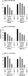
Antigen presentation assays using IE antigen–specific CD8 + T cell clones. Clones specific for the BZLF1-derived EPL (HLA-B*3501 restricted) and RAK (HLA-B*0801 restricted) epitopes and the BRLF1- derived YVL (HLA-A*0201 restricted) epitope were tested in ELISPOT assays against pairs of BZLF1+ and BZLF1− LCLs with the relevant HLA class I–restricting allele (where 5, 10, and 5% of the BZLF1+ LCL cells, respectively, were in lytic cycle) and on the BZLF1-LCL in which 1, 2, or 5% of cells had been preexposed to the epitope peptide. Results are shown as the number of spot-forming cells per well, and the error bars represent 1 SD. Data from the two EPL-specific clones are representative of results obtained on the only available HLA-B*3501–positive LCL pair. Data from the two RAK-specific clones and the two YVL-specific clones are in each case representative of results obtained from three clones of each specificity tested on three different LCL pairs.
Figure 6.
Figure 6.
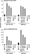
Antigen presentation assays using E antigen–specific CD8 + T cell clones. Clones specific for the BMLF1-derived GLC (HLA-A*0201 restricted), the BMRF1-derived TLD (HLA-A*0201 restricted) and the BALF2-derived ARYA (HLA-B*2705 restricted) epitopes were assayed as in Fig. 5; in each case, 5% of the BZLF1+ LCL cells were in lytic cycle. Data from the two GLC-specific and TLD-specific clones are each representative of results obtained for three epitope-specific clones tested on two different HLA-A*0201–positive LCL pairs, and data from the two ARYA-specific clones are representative of results obtained from four such clones on two different HLA-B*2705–positive LCL pairs.
Figure 7.
Figure 7.
Antigen presentation assays using L antigen–specific CD8 + T cell clones. Clones specific for the BALF4-derived FLD (HLA-A*0201 restricted) and the BILF2-derived RRRK (HLA-B*2705 restricted) epitopes were assayed as in Fig. 5; for each set of effectors, 4% of the BZLF1+ LCL cells were in late lytic cycle. These assays also included the ACV-treated BZLF1+ LCL (with no cells in late lytic cycle) as an additional target (ACV). Data from the FLD-specific clones are representative of results from three such clones tested on two different HLA-A*0201–positive LCL pairs, and data from the two RRRK-specific clones are representative of results obtained on two different HLA-B*2705–positive LCL pairs.
Figure 8.
Figure 8.
Comparative antigen presentation and functional avidity assays using representative CD8 + T cell clones specific for IE, E, and L protein–derived epitopes. (A) HLA-A*0201–restricted epitopes, YVL from the IE protein BRLF1, GLC from the E protein BMLF1, TLD from the E protein BMRF1, and FLD from the L protein BALF4, and (B) HLA-B*2705– restricted epitopes, ARYA from the E protein BALF2 and RRRK from the L protein BILF2. In each case, the top half of the figure shows the results of peptide titration assays (performed on a peptide-loaded BZLF1− LCL) plotting number of spot-forming cells against peptide concentration; this allows functional avidity of the CD8+ T cell clone to be determined as the peptide concentration mediating 50% optimal recognition. The bottom half of the figure in each case shows the results of antigen presentation assays on a pair of BZLF1+ and BZLF1− LCL target cells (where 5% of the BZLF1+ LCL cells were in lytic cycle) and on the BZLF1− LCL in which 5% cells had been loaded with epitope peptide. Note that these results are from a single experiment in which all 7 clones were tested simultaneously on the same BZLF1+ and BZLF1− LCL pair derived from a HLA-A*0201, B*2705-positive donor, and titrated on the same BZLF1− LCL loaded with cognate peptide. Data are expressed as in Fig. 5.

References

    1. Doherty, P.C., and J.P. Christensen. 2000. Accessing complexity: the dynamics of virus-specific T cell responses. Annu. Rev. Immunol. 18:561–592. - PubMed
    1. McMichael, A.J., and R.E. Phillips. 1997. Escape of human immunodeficiency virus from immune control. Annu. Rev. Immunol. 15:271–296. - PubMed
    1. Klenerman, P., V. Cerundolo, and P.R. Dunbar. 2002. Tracking T cells with tetramers: new tales from new tools. Nat. Rev. Immunol. 2:263–272. - PubMed
    1. Yewdell, J.W., and J.R. Bennink. 1999. Immunodominance in major histocompatibility complex class I-restricted T lymphocyte responses. Annu. Rev. Immunol. 17:51–58. - PubMed
    1. Cresswell, P., N. Bangia, T. Dick, and G. Diedrich. 1999. The nature of the MHC class I peptide loading complex. Immunol. Rev. 172:21–28. - PubMed

Publication types

MeSH terms

Substances