. 2005 Feb 7;201(3):453-63.
doi: 10.1084/jem.20041672. Epub 2005 Jan 31.

Synergistic role of micronemal proteins in Toxoplasma gondii virulence

Affiliations

Synergistic role of micronemal proteins in Toxoplasma gondii virulence

Odile Cérède et al. J Exp Med. .

Abstract

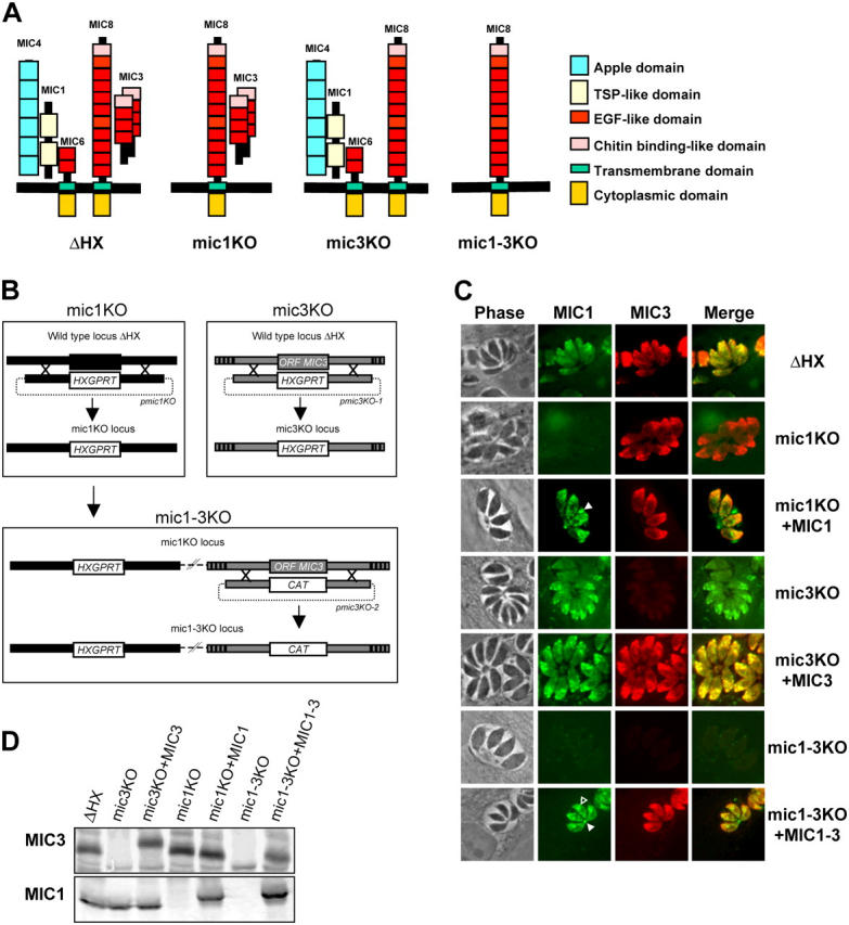
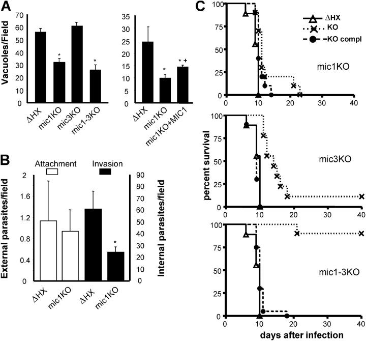
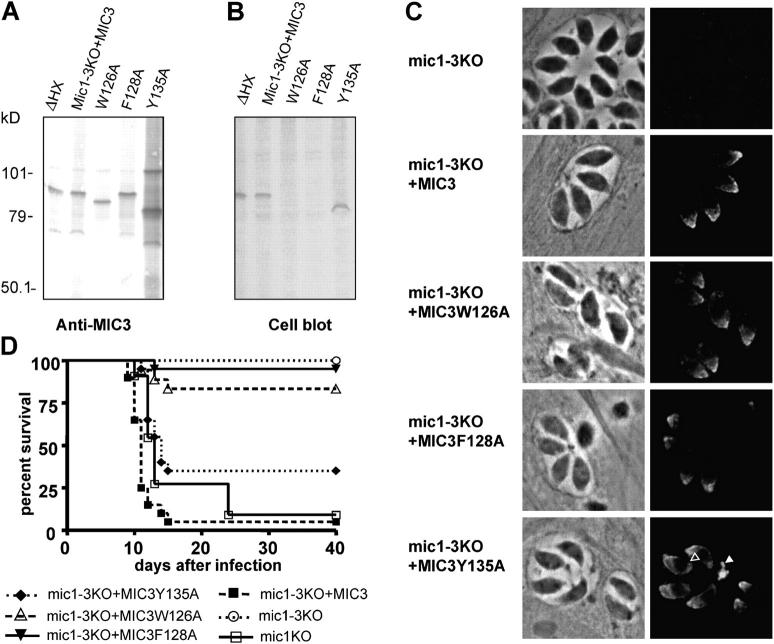
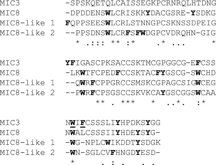
Apicomplexan parasites invade cells by a unique mechanism involving discharge of secretory vesicles called micronemes. Microneme proteins (MICs) include transmembrane and soluble proteins expressing different adhesive domains. Although the transmembrane protein TRAP and its homologues are thought to bridge cell surface receptors and the parasite submembranous motor, little is known about the function of other MICs. We have addressed the role of MIC1 and MIC3, two soluble adhesins of Toxoplasma gondii, in invasion and virulence. Single deletion of the MIC1 gene decreased invasion in fibroblasts, whereas MIC3 deletion had no effect either alone or in the mic1KO context. Individual disruption of MIC1 or MIC3 genes slightly reduced virulence in the mouse, whereas doubly depleted parasites were severely impaired in virulence and conferred protection against subsequent challenge. Single substitution of two critical amino acids in the chitin binding-like (CBL) domain of MIC3 abolished MIC3 binding to cells and generated the attenuated virulence phenotype. Our findings identify the CBL domain of MIC3 as a key player in toxoplasmosis and reveal the synergistic role of MICs in virulence, supporting the idea that parasites have evolved multiple ligand-receptor interactions to ensure invasion of different cells types during the course of infection.


Figures

Figure 1.
Figure 1.
Disruption of MIC1 and MIC3 genes and genetic complementation. (A) Schematic representations of the MIC1/4/6 and MIC3/8 complexes in micronemes of the ΔHX strain and the KO strains constructed in this study. In mic1KO (and mic 1-3KO), MIC1 is ablated, whereas MIC4 and MIC6 are mistargeted, resulting in a triple deletion of the proteins in micronemes. (B) Schematic drawing of the KO procedures described in Results. (C) IF of ΔHX (wild-type), MIC KOs, and complemented parasites. In wild-type, polyclonal anti-MIC3 (red) and mAb anti-MIC1 (green) show a punctuate fluorescence pattern within the apical complex, characteristic of microneme staining. Anti-MIC1 does not label mic1KO and mic1-3KO, whereas mic3KO and mic1-3KO do not react with anti-MIC3. Reexpression of MIC3 in mic3KO and mic1-3KO fully restored microneme labeling, whereas reexpression of MIC1 in mic1KO+MIC1 and in mic1-3KO+MIC1-3 led to some parasitophorous vacuole (closed arrowhead) and perinuclear (open arrowhead) labeling in addition to microneme staining. (D) Western blot analysis of MIC1 and MIC3 expression. On the top, the membrane was probed with anti-MIC3, and on the bottom, it was probed with anti-MIC1. The mobility shift observed for MIC3 in mic3KO+MIC3 is consistent with the addition of the Ty-1 epitope tag in this strain. Note the mobility shift of MIC1 in both complemented strains due to the addition of an myc epitope tag.
Figure 2.
Figure 2.
Phenotypes of MIC KO parasites. (A) Deletion of the MIC1 gene reduces invasion in HFF cells. Confluent monolayers of HFF cells were incubated with tachyzoites, and invasion was assessed as described in Materials and methods. Results are presented as the number of vacuoles per field. Data are mean values ± SEM determined by quadruple assays (10 fields per assay) performed in five separate experiments. Asterisk and cross indicate a significant difference (P < 0.05, two-tailed Student's t test) compared with ΔHX and mic1KO, respectively. (B) Differentiation of invaded parasites from attached parasites with a red/green invasion assay. Data are mean values ± SEM of parasites by field determined by triplicate assay (10 fields per assay) performed in four separate experiments. The asterisk indicates a statistically significant difference compared with ΔHX. (C) Single deletion of MIC1 or MIC3 gene slightly reduced virulence in mice, whereas the double KO severely impaired virulence. 20 tachyzoites of the indicated strains were injected i.p. into OF1 male mice (n = 10), and mouse survival was monitored daily for 40 d. The mic1KO, mic3KO, and mic1-3KO were statistically less virulent than wild-type (P < 0.002, P < 0.0001, and P < 0.0001, respectively, by Logrank test), and the double deletion significantly reduced the virulence compared with the single deletion (P < 0.0001).
Figure 3.
Figure 3.
Identification of critical residues in the CBL domain of MIC3. (A) Representation of conserved residues in the CB type 1 domain (PROSITE PDOC00025, CB type 1 domain signature and profile) and the CBL domain of T. gondii MIC3 (CAB56644). The topological arrangement of the four disulfide bonds is indicated by brackets. Conserved residues of the binding site are in red letters and well-conserved positions are in blue letters. C, cysteine; S, serine; P, proline; X, any amino acid; Φ, aromatic amino acid. All amino acids are contiguous, but the sequences are represented with a carriage return before each cysteine. (B) IF of mutated MIC3 proteins in transfected cells. BHK-21 cells were transfected with plasmids allowing expression of MIC3 or mutated MIC3 tagged with V5 epitope and coexpression of the green fluorescent protein. Cytoplasmic fluorescence of green fluorescent protein allows the monitoring of transfected cells. MIC3 was visualized with mAb anti-V5 (red) after permeabilization (Perm, intracellular MIC3) or without permeabilization (No perm, surface-bound MIC 3). Secretion was monitored by Western blotting. (d, e, and f). Equivalent fractions of cells (pellet) and corresponding supernatants (SN) collected 18 h after transfection were probed with mAb anti-V5. Three phenotypes were obtained: the recombinant protein is retained in the secretory pathway (exemplified by Y141 in a and d); the protein is secreted and binds to the surface of transfected cells (exemplified by R-MIC3 in b and e); and the protein is secreted but does not bind to the surface of transfected cells (exemplified by F128A in c and f). Adhesive proteins show a stronger signal in the pellet, resulting from their surface association, in contrast to nonadhesive R-MIC3 where a stronger signal is obtained in the supernatant.
Figure 4.
Figure 4.
The cell binding property of MIC3 is crucial for virulence. Single amino acid mutations were generated in the MIC3 gene and transfected in the mic1-3KO mutant. (A) Western blot of transgenic parasites under nonreducing conditions. The nitrocellulose membrane was probed with mAb anti-MIC3. (B) Cell blot analysis of transgenic parasites. A duplicate nitrocellulose membrane was incubated with Mode-K cells, washed, and bound cells were stained with amidoblack. Cells do not bind to W126A and F128A. (C) Phase contrast and IF of intracellular tachyzoites showing expression and microneme localization of MIC3 in the single amino acid mutants. Y135A in mic1-3KO also accumulates in the parasitophorous vacuole (closed arrowhead) and in the perinuclear area (open arrowhead). (D) Virulence of transgenic parasites. 20 tachyzoites of the indicated strains were injected i.p. into OF1 male mice (n = 20) that were monitored for 42 d. The mic1-3KO and the mutants complemented with W126A and F128A are markedly impaired in virulence and were not statistically different from mic1-3KO (Logrank test), whereas complementation with MIC3 or Y135A restored totally or partially the mic1KO phenotype. Mic1-3KO+MIC3Y135A was statistically more virulent than mic1-3KO (P < 0.0009) but was significantly less virulent than mic1-3KO+MIC3 (P < 0.0001).
Figure 5.
Figure 5.
Alignment of the amino acid sequences of the CBL domain of MIC3, MIC8 (AAK19757), and of putative transmembrane MICs closely related to MIC8 (named MIC8–like 1 and MIC8–like 2) present in the T. gondii genome resource (reference 29). Aromatic acids are shown in bold and those involved in cell binding properties of MIC3 are underlined.

References

    1. Menard, R. 2001. Gliding motility and cell invasion by Apicomplexa: insights from the Plasmodium sporozoite. Cell. Microbiol. 3:63–73. - PubMed
    1. Soldati, D., J.F. Dubremetz, and M. Lebrun. 2001. Microneme proteins: structural and functional requirements to promote adhesion and invasion by the apicomplexan parasite Toxoplasma gondii. Int. J. Parasitol. 31:1293–1302. - PubMed
    1. Tomley, F.M., and D.S. Soldati. 2001. Mix and match modules: structure and function of microneme proteins in apicomplexan parasites. Trends Parasitol. 17:81–88. - PubMed
    1. Matuschewski, K., A.C. Nunes, V. Nussenzweig, and R. Menard. 2002. Plasmodium sporozoite invasion into insect and mammalian cells is directed by the same dual binding system. EMBO J. 21:1597–1606. - PMC - PubMed
    1. Kappe, S., T. Bruderer, S. Gantt, H. Fujioka, V. Nussenzweig, and R. Menard. 1999. Conservation of a gliding motility and cell invasion machinery in apicomplexan parasites. J. Cell Biol. 147:937–944. - PMC - PubMed

Publication types

MeSH terms

Substances