. 2005 Feb;25(4):1258-71.
doi: 10.1128/MCB.25.4.1258-1271.2005.

Nucleophosmin (B23) targets ARF to nucleoli and inhibits its function

Affiliations

Nucleophosmin (B23) targets ARF to nucleoli and inhibits its function

Chandrashekhar Korgaonkar et al. Mol Cell Biol. 2005 Feb.

Abstract

The ARF tumor suppressor is a nucleolar protein that activates p53-dependent checkpoints by binding Mdm2, a p53 antagonist. Despite persuasive evidence that ARF can bind and inactivate Mdm2 in the nucleoplasm, the prevailing view is that ARF exerts its growth-inhibitory activities from within the nucleolus. We suggest ARF primarily functions outside the nucleolus and provide evidence that it is sequestered and held inactive in that compartment by a nucleolar phosphoprotein, nucleophosmin (NPM). Most cellular ARF is bound to NPM regardless of whether cells are proliferating or growth arrested, indicating that ARF-NPM association does not correlate with growth suppression. Notably, ARF binds NPM through the same domains that mediate nucleolar localization and Mdm2 binding, suggesting that NPM could control ARF localization and compete with Mdm2 for ARF association. Indeed, NPM knockdown markedly enhanced ARF-Mdm2 association and diminished ARF nucleolar localization. Those events correlated with greater ARF-mediated growth suppression and p53 activation. Conversely, NPM overexpression antagonized ARF function while increasing its nucleolar localization. These data suggest that NPM inhibits ARF's p53-dependent activity by targeting it to nucleoli and impairing ARF-Mdm2 association.


Figures

FIG. 1.
FIG. 1.
ARF associates with nucleolar phosphoproteins NPM and nucleolin. Narf6 cells expressing IPTG-inducible human ARF were treated with IPTG (+) or not (−) for 2 days. (A) Cells were labeled with 32P- labeled inorganic phosphate, and ARF complexes were immunoprecipitated (IP) with two different antibodies against human ARF (from Novus and Sigma). ARF and its associated phosphoproteins were detected by autoradiography and immunoblotting, as indicated. Coincident detection of ARF (indicated by @) and associated human Mdm2 (Hdm2; #), NPM (**), and nucleolin (*) is denoted by symbols on the autoradiogram and Western blots. (B) Association between phosphorylated C23/nucleolin with ARF and NPM was demonstrated in 32P-labeled Narf6 cells by immunoprecipitation with ARF and NPM antibodies followed by Western blotting for nucleolin, as described for panel A. (C) Colocalization of ARF and NPM in Narf6 cells, indicated by their overlapping staining patterns (merge), was examined by confocal microscopy after immunofluorescent staining with antibodies to ARF and NPM. Individual cells are shown by phase contrast microscopy. (D) Reciprocal immunoprecipitation-Western blotting was performed with Narf6 cell lysates with control IgG, ARF, and NPM antibodies for immunoprecipitation, followed by immunoblotting with ARF and NPM antibodies. Expression levels of ARF and NPM in the cell lysates (10% input into the immunoprecipitations) were also examined.
FIG. 2.
FIG. 2.
ARF associates with NPM independently of p53 and cell growth status. Complexes between endogenous ARF and NPM in proliferating 10-1 cells (A) or in mouse NIH 3T3 fibroblasts infected with retroviruses encoding vector (lanes V) or ARF (lanes A) (B) were detected by reciprocal immunoprecipitation-Western blotting with ARF and NPM antibodies, as denoted. IgG served as a negative control. Representative histograms, obtained from flow cytometric analysis of propidium iodide-stained nuclei, show the cell cycle distributions for 10-1 cells (panel A) and ARF-arrested NIH 3T3 cells (panel B). The relative percentage of cells in S phase is denoted and highlighted in black.
FIG. 3.
FIG. 3.
Nucleolar localization and Mdm2 binding domains of ARF mediate association with NPM. (A) In vivo complexes between endogenous NPM and ectopically expressed forms of mouse ARF were examined by reciprocal immunoprecipitation-Western blotting in COS or NIH 3T3 cells, as denoted. COS cells were transfected with empty vector, wild-type ARF, or the indicated deletion mutants, whereas 3T3 cells were infected with viruses encoding vector or various forms of ARF. The ARF double mutant (DM) lacks residues 1 to 14 and 26 to 37. (B) GST or GST-NPM proteins were mixed with equivalent amounts of 35S-labeled in vitro-translated mouse ARF or the indicated mutants. Specific in vitro binding between GST-NPM and various forms of ARF was detected by autoradiography (exposures are identical since all samples were analyzed on a single gel). The addition of equivalent levels of GST and GST-NPM proteins to the reactions was confirmed by Coomassie blue staining.
FIG. 4.
FIG. 4.
ARF associates with the nucleolar targeting domain of NPM. (A) A schematic of NPM depicts its N-terminal homo-oligomerization domain (HoD; residues 1 to 117), nuclear localization signal (NLS), and C-terminal heterodimerization (HeD; 187 to 259) and nucleic acid binding domains (NBD; 259 to 295). (B) For in vivo binding analyses, COS cells were transfected (+) or not (−) with plasmids encoding wild-type human ARF plus empty vector containing an HA tag (HA Vec), HA-tagged wild-type NPM (HA.NPM), or the indicated HA-tagged mutants of NPM. HA-NPM proteins were immunoprecipitated with HA-agarose and detected by immunoblotting with HA antibodies. Association between the HA-NPM proteins with ARF and endogenous NPM (* indicates exogenous HA-NPM) or nucleolin (C23) was detected by Western blotting. (C) In vitro binding of radiolabeled human ARF to GST, GST-wild-type NPM (WT), or various GST-NPM mutants (NPM residues fused to GST are indicated) was detected by autoradiography. Coomassie blue staining of the gel demonstrated equivalent levels of input GST proteins.
FIG. 5.
FIG. 5.
NPM contributes to the nucleolar localization of ARF. (A) Immunoblotting demonstrated stable small hairpin RNA-mediated knockdown of NPM in two U2OS-derived clonal populations (kd.1 and kd.2) compared to normal levels of NPM and overexpressed HA-NPM (*) in control (CON) cells. Tubulin served as a loading control. (B) Immunoblotting of NPM, ARF, and tubulin in NIH 3T3 and p21−/− mouse embryo fibroblasts infected with the vector control (−) or ARF (+) retrovirus and in Narf6 cells treated with IPTG (+) or not (−). ARF induced complete growth arrest in each cell type (data not shown). (C) Empty vector (Vec) or plasmids encoding wild-type human ARF were transfected into kd.1 or control cells, and ARF localization was assessed by immunofluorescence. Nuclei were visualized by DAPI. (D) Nucleolar integrity remains intact in NPM knockdown cells, as indicated by staining for the nucleolar protein fibrillarin.
FIG. 6.
FIG. 6.
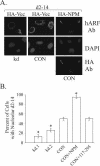
Dose-dependent effect of NPM on ARF localization. (A) Human ARF mutant d2-14 was expressed with empty HA-vector or HA-NPM in kd.1 or control cells, as indicated. Localization of the ARF mutant was examined by immunofluorescence. (B) Quantification of the data from panel A, showing a dose-dependent increase of d2-14 nucleolar localization in cells expressing increasing levels of NPM. Data were averaged from at least three independent experiments in which 100 or more cells were counted per sample. Error bars indicate the standard deviation. Asterisks denote statistically significant differences (P values all less than 0.0013) between the indicated samples and control cells, as determined by a paired, two-tailed Student t test.
FIG. 7.
FIG. 7.
NPM inhibits ARF function. (A) Plasmids containing human Mdm2 (Hdm2) and human ARF were cotransfected into control (C) and knockdown (kd) cells, and their expression (50 μg/lane) was detected by immunoblotting. ARF-human Mdm2 complexes were detected by reciprocal immunoprecipitation-Western blotting (from 500 μg of lysates) with ARF and human Mdm2 antibodies. (B) Increasing amounts of NPM plasmid (0, 2.5, 5, or 10 μg) were introduced into U2OS cells with constant levels of ARF construct (1 μg), and the relative transcriptional activity of p53 was measured with a p53-luciferase reporter assay. Data were averaged from two independent experiments. (C) Bromodeoxyuridine (BrdU) incorporation was measured in knockdown (kd) cells expressing human ARF (black bar), control (CON) cells expressing human ARF (hatched bar), or control cells expressing human ARF plus NPM (open bar). Both cell types were transfected with empty vector as the control and incorporated equivalent amounts of bromodeoxyuridine (knockdown = 89.5%, control = 90.5%). At least 100 ARF-positive cells were scored in each experiment, and error bars represent standard deviations for at least three independent experiments. The indicated P values denote statistically significant differences between ARF activities under the different conditions. (D) Control and knockdown cells were transfected with empty vector or plasmids expressing ARF or NPM, and equivalent amounts of lysate were analyzed by Western blotting for expression of ARF, NPM (* denotes exogenous HA-NPM), p53, human Mdm2, p21, and C23/nucleolin (loading control), as indicated. Matched exposures are given since samples were electrophoresed on separate gels.
FIG. 8.
FIG. 8.
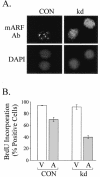
ARF mutant acquires growth-inhibitory activity upon NPM knockdown. (A) The mouse ARF mutant D21-25 was expressed in NPM knockdown (kd) or control (CON) cells, and its localization was detected by immunofluorescence and confocal microscopy. Nuclei were visualized by DAPI staining. (B) Bromodeoxyuridine (BrdU) incorporation was determined in knockdown and control cells transfected with empty vector (V; open bar) or the D21-25 ARF mutant (A; gray bar). Data were averaged from two independent experiments where at least 80 cells were scored for each condition.
FIG. 9.
FIG. 9.
Models of ARF and NPM coregulation. (A) ARF and NPM exist in a negative regulatory feedback loop. We discovered that NPM-mediated nucleolar targeting inhibits several ARF functions (human Mdm2 [Hdm2] binding, p53 activation, and growth suppression) despite enhancing ARF expression, which correlates with reduced ARF degradation in nucleoli (32, 64). These findings complement previous observations that ARF promotes ubiquitin-dependent degradation of NPM and inhibits NPM-mediated rRNA processing (2, 25, 72). (B) Model showing how NPM negatively regulates p53-dependent ARF signaling. ARF is not expressed in most normal cells, where human Mdm2 maintains low levels of p53 in the nucleoplasm (shaded gray) by enhancing its degradation in the cytoplasm (indicated by arrow). Oncogenic stimuli induce both ARF and NPM; initially high levels of NPM sequester ARF in nucleoli, thereby enabling continued cell division (incipient cancer cell). Over time, NPM stabilizes ARF, which in turn promotes the ubiquitination and downregulation of NPM. This facilitates release of a fraction of ARF into the nucleoplasm, where it associates with human Mdm2, leading to p53 activation and suppression of cell growth (arrested or dying cell). In conjunction with other genetic changes (Δs), the cancer-associated upregulation of NPM could make it impervious to negative regulation by ARF, promoting unrestrained proliferation in the presence of ARF (cancer cell).

References

    1. Bertwistle, D., F. Zindy, C. J. Sherr, and M. F. Roussel. 2004. Monoclonal antibodies to the mouse p19ARF tumor suppressor protein. Hybridoma Hybridomics 23:293-300. - PubMed
    1. Bertwistle, D., M. Sugimoto, and C. J. Sherr. 2004. Physical and functional interactions of the Arf tumor suppressor protein with nucleophosmin/B23. Mol. Cell. Biol. 24:985-996. - PMC - PubMed
    1. Biggiogera, M., K. Burki, S. H. Kaufmann, J. H. Shaper, N. Gas, F. Amalric, and S. Fakan. 1990. Nucleolar distribution of proteins B23 and nucleolin in mouse preimplantation embryos as visualized by immunoelectron microscopy. Development 110:1263-1270. - PubMed
    1. Borer, R. A., C. F. Lehner, H. M. Eppenberger, and E. A. Nigg. 1989. Major nucleolar proteins shuttle between nucleus and cytoplasm. Cell 56:379-390. - PubMed
    1. Carnero, A., J. D. Hudson, C. M. Price, and D. H. Beach. 2000. p16(INK4A) and p19ARF act in overlapping pathways in cellular immortalization. Nat. Cell Biol. 2:148-155. - PubMed

Publication types

MeSH terms