. 2005 Feb;25(4):1354-66.
doi: 10.1128/MCB.25.4.1354-1366.2005.

Control of mitochondrial transcription specificity factors (TFB1M and TFB2M) by nuclear respiratory factors (NRF-1 and NRF-2) and PGC-1 family coactivators

Affiliations

Control of mitochondrial transcription specificity factors (TFB1M and TFB2M) by nuclear respiratory factors (NRF-1 and NRF-2) and PGC-1 family coactivators

Natalie Gleyzer et al. Mol Cell Biol. 2005 Feb.

Abstract

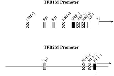
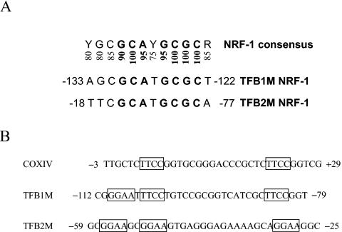
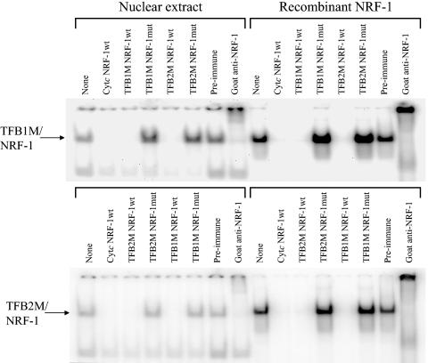
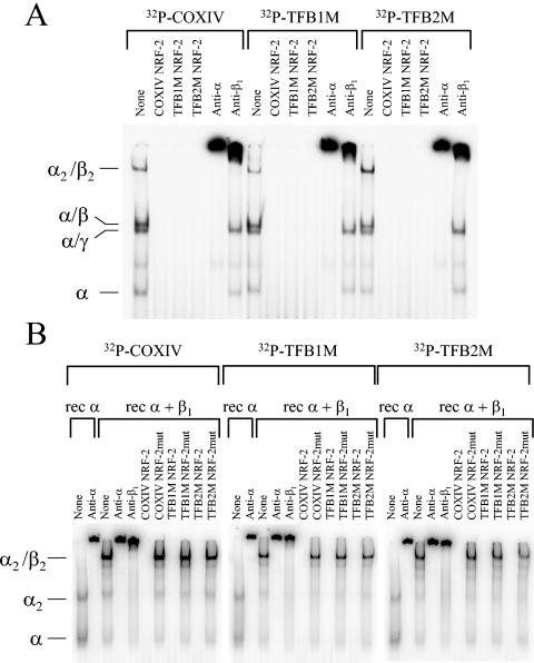
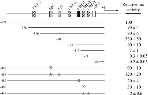
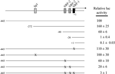
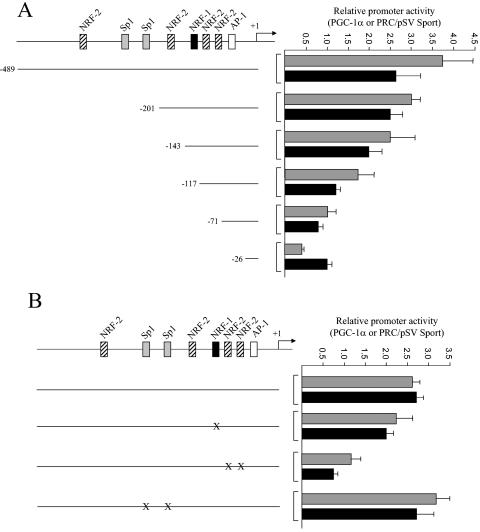
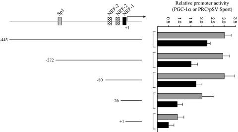
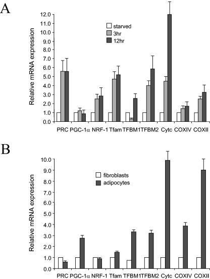
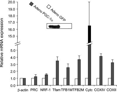
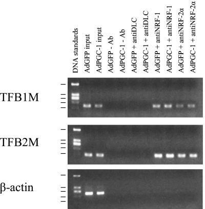
In vertebrates, mitochondrial DNA (mtDNA) transcription is initiated bidirectionally from closely spaced promoters, HSP and LSP, within the D-loop regulatory region. Early studies demonstrated that mtDNA transcription requires mitochondrial RNA polymerase and Tfam, a DNA binding stimulatory factor that is required for mtDNA maintenance. Recently, mitochondrial transcription specificity factors (TFB1M and TFB2M), which markedly enhance mtDNA transcription in the presence of Tfam and mitochondrial RNA polymerase, have been identified in mammalian cells. Here, we establish that the expression of human TFB1M and TFB2M promoters is governed by nuclear respiratory factors (NRF-1 and NRF-2), key transcription factors implicated in mitochondrial biogenesis. In addition, we show that NRF recognition sites within both TFB promoters are required for maximal trans activation by the PGC-1 family coactivators, PGC-1alpha and PRC. The physiological induction of these coactivators has been associated with the integration of NRFs and other transcription factors in a program of mitochondrial biogenesis. Finally, we demonstrate that the TFB genes are up-regulated along with Tfam and either PGC-1alpha or PRC in cellular systems where mitochondrial biogenesis is induced. Moreover, ectopic expression of PGC-1alpha is sufficient to induce the coordinate expression of all three nucleus-encoded mitochondrial transcription factors along with nuclear and mitochondrial respiratory subunits. These results support the conclusion that the coordinate regulation of nucleus-encoded mitochondrial transcription factors by NRFs and PGC-1 family coactivators is essential to the control of mitochondrial biogenesis.


Figures

FIG. 1.
FIG. 1.
Schematic representation of hTFB1M and hTFB2M promoters. The hTFB1M promoter was identified by aligning the CGI-75 cDNA (accession number NP_057104) corresponding to hTFB1M (20) with the human genomic sequence from chromosome 6 encoding this gene (accession number AL139101). The hTFB2M promoter was identified by aligning the human cDNA (accession number NM_022366) with the human genomic clone from chromosome 1 (accession number AL356583). The cis-acting elements, denoted by labeled rectangular boxes, were initially identified by the Match internet search tool and by visual inspection of the sequence.
FIG. 2.
FIG. 2.
Comparison of hTFB NRF-1 and NRF-2 sites. (A) NRF-1 sites from hTFB1M and hTFB2M are compared to the consensus. Numbers below the consensus represent the percent representation of the nucleotide at that position in over 20 functional NRF-1 sites. Highly invariant nucleotides are in boldface. (B) Comparison of tandemly arranged NRF-2 sites in the TFB promoters to those present in the COXIV promoter in which they were first identified. The GGAA core motifs are boxed. Numbers adjacent to the sequences denote the nucleotide positions relative to the mRNA 5′ ends.
FIG. 3.
FIG. 3.
Specific binding of NRF-1 to recognition sites within the hTFB1M and hTFB2M promoters. Radiolabeled synthetic oligonucleotides containing either the TFB1M or TFB2M NRF-1 site were bound to either crude nuclear extracts or purified recombinant NRF-1, and the complexes (indicated by the arrows) were separated by gel electrophoresis. The unlabeled competitor oligonucleotide or the supershifting antiserum added to the binding reaction is indicated above each lane.
FIG. 4.
FIG. 4.
Specific binding of NRF-2 to recognition sites within the hTFB1M and hTFB2M promoters. (A) Radiolabeled synthetic oligonucleotides containing COXIV, hTFB1M, or hTFB2M recognition sites were bound to an aliquot of partially purified nuclear extract that was eluted from heparin agarose at 0.25 M NaCl. The indicated DNA-protein complexes were separated by gel electrophoresis. The unlabeled competitor oligonucleotide or the supershifting antiserum added to the binding reactions is indicated above each lane. (B) The same radiolabeled oligonucleotides as in panel A were bound to either recombinant NRF-2α (rec α) or a mixture of recombinant NRF-2α and NRF-2β1 (rec α + β1). The indicated DNA-protein complexes were separated by gel electrophoresis. The unlabeled competitor oligonucleotide or the supershifting antiserum added to the binding reactions is indicated above each lane.
FIG. 5.
FIG. 5.
Mutational analysis of the hTFB1M promoter region. A series of 5′ deletions designed to progressively remove putative cis-acting elements was analyzed by gene transfection. The normalized luciferase activity obtained from the promoter fragment containing 489 nucleotides of 5′-flanking DNA (−489) was designated as 100%. Point mutations represented by X were introduced into the −489 promoter as described under Materials and Methods. The activities of all of the mutated promoters were expressed as a percentage of that obtained from the −489 promoter. Numbers represent the average ± standard error for three separate determinations.
FIG. 6.
FIG. 6.
Mutational analysis of the hTFB2M promoter region. A series of 5′ deletions designed to progressively remove putative cis-acting elements was analyzed by gene transfection. The normalized luciferase activity obtained from the promoter fragment containing 443 nucleotides of 5′-flanking DNA (−443) was designated as 100%. Point mutations represented by X were introduced into the −443 promoter as described under Materials and Methods. The activities of all of the mutated promoters were expressed as a percentage of that obtained from the −443 promoter. Numbers represent the average ± standard error for three separate determinations.
FIG. 7.
FIG. 7.
trans activation of the hTFB1M promoter and its mutated derivatives by PGC-1α and PRC. (A) The hTFB1M promoter deletions shown in Fig. 5 were assayed for trans activation by PGC-1α (gray bars) and PRC (black bars) by cotransfection with vectors expressing each coactivator. Values are the average fold activation for three separate determinations ± standard error measured relative to the pSVSport negative control. (B) Same as panel A except that the indicated point mutations were assayed for trans activation by the coactivators.
FIG. 8.
FIG. 8.
trans activation of the hTFB2M promoter and its mutated derivatives by PGC-1α and PRC. The hTFB2M promoter deletions shown in Fig. 6 were assayed for trans activation by PGC-1α (gray bars) and PRC (black bars) by cotransfection with vectors expressing each coactivator. Values are the average fold activation for three separate determinations ± standard error measured relative to the pSVSport negative control.
FIG. 9.
FIG. 9.
Expression of hTFB genes compared to that of a collection of regulatory and structural genes involved in mitochondrial biogenesis. (A) Gene expression was monitored during serum stimulation of quiescent BALB/3T3 fibroblasts by quantitative real-time PCR. The battery of genes examined represented nuclear regulatory factors (PRC, PGC-1α, and NRF-1), mitochondrial transcription and replication factors (Tfam, TFB1M, and TFB2M), and nucleus (Cyt c and COXIV)- and mitochondrion (COXII)-encoded respiratory subunits. Relative steady-state mRNA levels were normalized to 18S rRNA as an internal control. (B) Relative mRNA expression was monitored during differentiation of fibroblasts to adipocytes for the same battery of genes as in panel A. Steady-state mRNA levels were normalized to rRNA and represent the average of at least three separate determinations with error bars denoting ± standard error.
FIG. 10.
FIG. 10.
Expression of hTFB genes in response to ectopic expression of PGC-1α. Expression of mRNAs was measured in response to production of PGC-1α from an adenovirus vector in C2C12 myoblasts compared to a GFP-producing control. The inset panel shows the expression of PGC-1α protein by immunoblotting. Steady-state mRNA levels determined 72 h postinfection were normalized to that of β-actin and represent the average of three separate determinations with error bars denoting ± standard error.
FIG. 11.
FIG. 11.
NRF-1 and NRF-2α occupancy of TFB1M and TFB2M promoters in vivo detected by ChIP. HeLa cells infected with Ad-GFP- or Ad-PGC-1-expressing virus were subjected to ChIP assays as described in Materials and Methods. Input lanes show the PCR product derived from chromatin prior to immunoprecipitation. Antibodies used for immunoprecipitation are indicated above each lane. Precipitated DNA was analyzed by semiquantitative PCR with primer sets specific for the TFB1M or TFB2M promoter or the control β-actin fragment as indicated. Sizes of the DNA standards indicated at the left are, from top to bottom, 1,207, 540, 400 (doublet), 275, and 166 bp.

Similar articles

Cited by

References

    1. Andersson, U., and R. C. Scarpulla. 2001. PGC-l-related coactivator, a novel, serum-inducible coactivator of nuclear respiratory factor 1-dependent transcription in mammalian cells. Mol. Cell. Biol. 21:3738-3749. - PMC - PubMed
    1. Andrews, N. C., and D. V. Faller. 1991. A rapid micropreparation technique for extraction of DNA-binding proteins from limiting numbers of mammalian cells. Nucleic Acids Res. 19:2499. - PMC - PubMed
    1. Antoshechkin, I., and D. F. Bogenhagen. 1995. Distinct roles for two purified factors in transcription of Xenopus mitochondrial DNA. Mol. Cell. Biol. 15:7032-7042. - PMC - PubMed
    1. Bogenhagen, D. F. 1996. Interaction of mtTFB and mtRNA polymerase at core promoters for transcription of Xenopus laevis mtDNA. J. Biol. Chem. 271:12036-12041. - PubMed
    1. Choi, Y. S., H. K. Lee, and Y. K. Pak. 2002. Characterization of the 5′-flanking region of the rat gene for mitochondrial transcription factor A (Tfam). Biochim. Biophys. Acta Gene Struct. Expr. 1574:200-204. - PubMed

Publication types

MeSH terms

Substances

LinkOut - more resources