. 2005 Jan 31;33(2):650-60.
doi: 10.1093/nar/gki211. Print 2005.

A highly specific phosphatase that acts on ADP-ribose 1''-phosphate, a metabolite of tRNA splicing in Saccharomyces cerevisiae

Affiliations

A highly specific phosphatase that acts on ADP-ribose 1''-phosphate, a metabolite of tRNA splicing in Saccharomyces cerevisiae

Neil P Shull et al. Nucleic Acids Res. .

Abstract

One molecule of ADP-ribose 1'',2''-cyclic phosphate (Appr>p) is formed during each of the approximately 500 000 tRNA splicing events per Saccharomyces cerevisiae generation. The metabolism of Appr>p remains poorly defined. A cyclic phosphodiesterase (Cpd1p) has been shown to convert Appr>p to ADP-ribose-1''-phosphate (Appr1p). We used a biochemical genomics approach to identify two yeast phosphatases that can convert Appr1p to ADP-ribose: the product of ORF YBR022w (now Poa1p), which is completely unrelated to other known phosphatases; and Hal2p, a known 3'-phosphatase of 5',3'-pAp. Poa1p is highly specific for Appr1p, and thus likely acts on this molecule in vivo. Poa1 has a relatively low K(M) for Appr1p (2.8 microM) and a modest kcat (1.7 min(-1)), but no detectable activity on several other substrates. Furthermore, Poa1p is strongly inhibited by ADP-ribose (K(I), 17 microM), modestly inhibited by other nucleotides containing an ADP-ribose moiety and not inhibited at all by other tested molecules. In contrast, Hal2p is much more active on pAp than on Appr1p, and several other tested molecules were Hal2p substrates or inhibitors. poa1-Delta mutants have no obvious growth defect at different temperatures in rich media, and analysis of yeast extracts suggests that approximately 90% of Appr1p processing activity originates from Poa1p.


Figures

Figure 1
Figure 1
Identification of two yeast ORFs associated with activity on Appr1p. (A) Assay of a genomic collection of pools of purified yeast GST–ORF fusion proteins. Pools of GST–ORF fusion proteins were assayed for Appr1p processing activity at 30°C for 4 h with 40 nM Ap*pr1p and products were resolved on PEI–cellulose TLC plates developed in 1 M sodium formate. a, buffer control. Products I, II and III are indicated. (B) Assay of sub-pools of plate 49 for activity on Appr1p. Sub-pools of GST–ORF fusion proteins from rows A–H and columns 1–12 of plate 49 were assayed with 3 nM Ap*pr1p at 30°C for 1 h, as indicated, and resolved as in (A). a, GST–ORF pool of plate 49.
Figure 2
Figure 2
Overexpression and purification of Poa1p and Hal2p from E.coli. (A) His6-YBR022w protein and Hal2p were purified as described in Materials and Methods from extracts of strains expressing His6-YBR022w protein (a–d) and His6-Hal2p (e–h), and analyzed by SDS–PAGE. a and e, 40 μg crude extract from strain expressing vector; b and f, 40 μg crude extract from strain expressing His6-YBR022w protein (b) or His6-Hal2p (f); c, d, g and h, 5 and 8 μg purified His6-YBR022w protein (c and d) or His6-Hal2p (g and h). (B) His6-YBR022w protein, Hal2p and CIP all act as a phosphatase on Appr1p. Reactions mixtures containing 19 nM Ap*pr1p substrate and 210 nM purified His6-YBR022w protein, 520 nM purified Hal2p or 56 fM CIP were incubated at 30°C, and reactions were stopped by spotting 2 μl to PEI–cellulose plates that were then resolved as in Figure 1. a and b, buffer control for 0 and 60 min; c–l, reactions containing YBR022w protein (c–g), Hal2p (h–l), or CIP (m–q) incubated for 0, 15, 30, 45 and 60 min. Migration of AMP (pA), ADP-ribose (Appr), and products I, II and III are indicated.
Figure 3
Figure 3
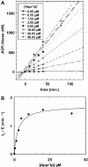
Kinetic parameters of Poa1p phosphatase for Appr1p. (A) Time courses of conversion of Appr1p to ADP-ribose at different substrate concentrations. Appr1p phosphatase activity was measured at 30°C for the times indicated, in reaction mixtures containing 21 nM Poa1p, 5–25 nM labeled Ap*pr1p and unlabeled Appr1p. Products were resolved as in Figure 2 and quantified, and data were plotted using KaleidaGraph software. (B) A Michaelis–Menten plot of the Appr1p phosphatase activity of Poa1p. Data from (A) were fit to a Michaelis–Menten curve using KaleidaGraph software.
Figure 4
Figure 4
The pH activity profile for Poa1p under kcat/KM conditions. Poa1p Appr1p phosphatase activity was measured at 30°C with 5 nM Poa1p and 25 nM Ap*pr1p at different pH values over a suitable time course, and rates were determined, as described in Materials and Methods. The following buffers were used: NaOAc, pH 4.0, 4.5, 4.7 and 5.0; Homo-PIPES, pH 4.7 and 5.0; MES, pH 5.4, 5.8, 6.0 and 6.2; Bis-Tris, pH 6.0, 6.6 and 7.0; HEPES, pH 7.0 and 7.5; and Tris–HCl, pH 7.5 and 8.0.
Figure 5
Figure 5
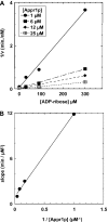
Determination of KI of ADP-ribose for Poa1p. (A) Plot of 1/v versus [ADP-ribose] for each of four Appr1p concentrations. Poa1p Appr1p phosphatase activity was measured at the indicated concentrations of Appr1p and ADP-ribose for a suitable time course as described in Figure 3, and rates were determined and plotted as indicated. (B) Plot of slope versus 1/[Appr1p]. Slopes were obtained for each curve in (A) above, and plotted versus 1/[Appr1p] as indicated. The slope of this plot (KM/Vmax KI) was used to determine the KI of ADP-ribose (17 μM), as described in Materials and Methods.
Figure 6
Figure 6
Comparison of Appr1p processing activity of crude extracts from wild-type cells and cells lacking Poa1p and/or Hal2p. Crude extracts derived from wild-type cells and poa1-Δ mutant, hal2-Δ mutant, or poa1-Δ, hal2-Δ double mutant cells were serially diluted 5-fold and assayed for Appr1p processing activity with 17 nM Ap*pr1p for 1 h at 30°C, as described in Materials and Methods. a–e, 16–0.04 μg wild-type extract; g–k, 23–0.04 μg hal2-Δ extract; l–p, 29 μg to 0.05 μg poa1-Δ extract; r–v, 21–0.03 μg poa1-Δ, hal2-Δ extract; f and q, buffer controls. Reactions were treated with phenol:chloroform and resolved by TLC as in Figure 1.

Similar articles

Cited by

References

    1. Peebles C.L., Gegenheimer P., Abelson J. Precise excision of intervening sequences from precursor tRNAs by a membrane-associated yeast endonuclease. Cell. 1983;32:525–536. - PubMed
    1. Ho C.K., Rauhut R., Vijayraghavan U., Abelson J. Accumulation of pre-tRNA splicing ‘2/3’ intermediates in a Saccharomyces cerevisiae mutant. EMBO J. 1990;9:1245–1252. - PMC - PubMed
    1. Trotta C.R., Miao F., Arn E.A., Stevens S.W., Ho C.K., Rauhut R., Abelson J.N. The yeast tRNA splicing endonuclease: a tetrameric enzyme with two active site subunits homologous to the archaeal tRNA endonucleases. Cell. 1997;89:849–858. - PubMed
    1. Phizicky E.M., Schwartz R.C., Abelson J. Saccharomyces cerevisiae tRNA ligase. Purification of the protein and isolation of the structural gene. J. Biol. Chem. 1986;261:2978–2986. - PubMed
    1. Phizicky E.M., Consaul S.A., Nehrke K.W., Abelson J. Yeast tRNA ligase mutants are nonviable and accumulate tRNA splicing intermediates. J. Biol. Chem. 1992;267:4577–4582. - PubMed

Publication types

MeSH terms

Substances