Comparative Study
. 2005 Feb 1:6:6.
doi: 10.1186/1471-2202-6-6.

The progressive nature of Wallerian degeneration in wild-type and slow Wallerian degeneration (WldS) nerves

Affiliations
Comparative Study

The progressive nature of Wallerian degeneration in wild-type and slow Wallerian degeneration (WldS) nerves

Bogdan Beirowski et al. BMC Neurosci. .

Abstract

Background: The progressive nature of Wallerian degeneration has long been controversial. Conflicting reports that distal stumps of injured axons degenerate anterogradely, retrogradely, or simultaneously are based on statistical observations at discontinuous locations within the nerve, without observing any single axon at two distant points. As axon degeneration is asynchronous, there are clear advantages to longitudinal studies of individual degenerating axons. We recently validated the study of Wallerian degeneration using yellow fluorescent protein (YFP) in a small, representative population of axons, which greatly improves longitudinal imaging. Here, we apply this method to study the progressive nature of Wallerian degeneration in both wild-type and slow Wallerian degeneration (WldS) mutant mice.

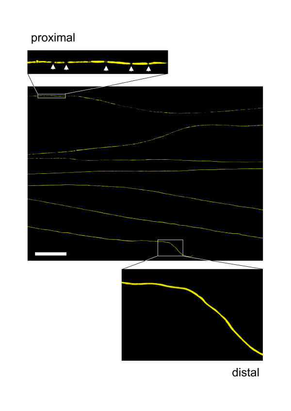
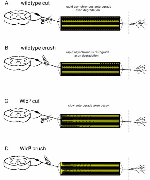
Results: In wild-type nerves, we directly observed partially fragmented axons (average 5.3%) among a majority of fully intact or degenerated axons 37-42 h after transection and 40-44 h after crush injury. Axons exist in this state only transiently, probably for less than one hour. Surprisingly, axons degenerated anterogradely after transection but retrogradely after a crush, but in both cases a sharp boundary separated intact and fragmented regions of individual axons, indicating that Wallerian degeneration progresses as a wave sequentially affecting adjacent regions of the axon. In contrast, most or all WldS axons were partially fragmented 15-25 days after nerve lesion, WldS axons degenerated anterogradely independent of lesion type, and signs of degeneration increased gradually along the nerve instead of abruptly. Furthermore, the first signs of degeneration were short constrictions, not complete breaks.

Conclusions: We conclude that Wallerian degeneration progresses rapidly along individual wild-type axons after a heterogeneous latent phase. The speed of progression and its ability to travel in either direction challenges earlier models in which clearance of trophic or regulatory factors by axonal transport triggers degeneration. WldS axons, once they finally degenerate, do so by a fundamentally different mechanism, indicated by differences in the rate, direction and abruptness of progression, and by different early morphological signs of degeneration. These observations suggest that WldS axons undergo a slow anterograde decay as axonal components are gradually depleted, and do not simply follow the degeneration pathway of wild-type axons at a slower rate.


Figures

Figure 1
Figure 1
After a latency period Wallerian degeneration following cut and crush injury starts abruptly in single axons and involves total fragmentation of axons within few hours A-D: Conventional fluorescence micrographs of a ~2.5 cm long peripheral nerve stump (sciatic-tibial nerve segment) wholemount preparation at the proximal (A) and distal site (B) 37 h after cut injury with few individual fluorescent axons broken into fragments. A small number of axons fragmented at the proximal (C) and distal site (D) of a peripheral nerve stump wholemount preparation could also be detected 40 h following crush injury. E-H: Conventional fluorescence micrographs of a ~2.5 cm long peripheral nerve stump (sciatic-tibial nerve segment) wholemount preparation at the proximal (E) and distal site (F) 42 h after cut injury with most YFP labelled axons fragmented. A similar picture with a majority of axons degenerated is evident at the proximal (G) and distal end (H) of a peripheral nerve stump wholemount preparation 44 h after crush injury. YFP fluorescence has been pseudo-coloured green with the applied imaging software (MetaVue, Universal Imaging Corporation). Magnification: 100 ×
Figure 2
Figure 2
Quantification of fluorescent axons in wholemount YFP-H peripheral nerve stumps after cut and crush injury at different time points. Depending on the extent of fragmentation, YFP positive axons from peripheral nerve stumps were assigned to the group "intact", "entirely fragmented", "fragmented with anterograde gradient" or "fragmented with retrograde gradient". The chart presents means and standard deviations. A: All partially fragmented axons that could be identified at the time points between 37 h and 42 h after cut injury were fragmented at the proximal end of the distal axon stump but not further distal, indicating an anterograde gradient of Wallerian degeneration ("fragmented with anterograde gradient"). A maximum of 9.3 % YFP positive axons with anterograde fragmentation appeared 40 h after cut injury. B: All partially fragmented axons that could be identified at the time points between 40 h and 44 h after crush injury were fragmented at their distal ends but not further proximal indicating a retrograde gradient of Wallerian degeneration ("fragmented with retrograde gradient"). A maximum of 7.2 % of YFP positive axons with retrograde fragmentation appeared 44 h after crush injury.
Figure 3
Figure 3
Wallerian degeneration proceeds in anterograde direction along individual axons after cut injury. Confocal composite picture showing seven consecutive lengths (from top to bottom in overview) of the proximodistal course of an individual YFP labelled axon within a distal nerve stump 40 h after transection demonstrating an anterograde progression of axon fragmentation. Note that this axon has fragmented in its proximal end (upper inset) but not in its distal end (lower inset). Axonal fragments are clearly demarcated by fluorescence interruptions (arrows in upper inset). YFP fluorescence has been pseudo-coloured yellow with the applied confocal imaging software (Biorad LaserSharp 2000). Scale bar: 500 μm
Figure 4
Figure 4
Wallerian degeneration proceeds in retrograde direction along individual axons after crush injury. Confocal composite picture showing seven consecutive lengths (from top to bottom in overview) of the proximodistal course of an individual YFP labelled axon within a peripheral nerve stump 44 h after crush injury displaying a retrograde progression of axon fragmentation. Note that this axon has fragmented in its distal end (lower inset) but not in its proximal end (upper inset). Axonal fragments are clearly demarcated by fluorescence interruptions (arrows in lower inset). YFP fluorescence has been pseudo-coloured yellow with the applied confocal imaging software (Biorad LaserSharp 2000). Scale bar: 500 μm
Figure 5
Figure 5
Wave front of Wallerian degeneration in a YFP labelled wild-type axon after crush lesion A: The partially degenerated axon that is bracketed was identified in a 44 h crushed wild-type nerve. All more distal regions of this axon are fragmented and all more proximal regions are intact (data not shown). B-D: higher magnification of this axon from (A) around the transition point between intact and fragmented regions. (D) shows the most proximal breakpoint in this nerve and the inferred retrograde direction of propagation of Wallerian degeneration. Immediately proximal to the breakpoint severe vacuolation occupies almost the entire axon thickness. Slightly further proximal in (C), there are also severe YFP negative vacuoles and fragmentation appears imminent at two points (asterisks). Further proximal still in (B), the degree of vacuolation decreases. YFP fluorescence has been pseudo-coloured green with the applied confocal imaging software (Zeiss LSM Software Release 3.2). Scale bars: 50 μm (A) and 10 μm (B, C, D)
Figure 6
Figure 6
Axonal fragmentation progresses asynchronously as a localised wave along individual axons in a anterograde or retrograde direction A-D: Graphs showing the number of axonal breaks along individual YFP labelled axons with anterograde gradient of fragmentation in relation to the distance in mm from the transection point 37 h (A), 40 h (B), 41 h (C) and 42 h (D) after cut lesion. Note that with increasing distance from the transection, axon lengths with marked fragmentation abruptly change into lengths with no or just a few axonal breaks, indicating that Wallerian degeneration progresses with a localised fragmentation wave front. Additionally note the variable localisation of the fragmentation wave front along different axons at one timepoint representing the asynchronity of Wallerian degeneration among the axon population. E-H: Graphs showing the number of axonal breaks along individual YFP labelled axons with retrograde gradient of fragmentation in relation to the distance in mm from the crush point 40 h (E), 42 h (F), 43 h (G) and 44 h (H) after crush lesion. Note that with increasing distance from the crush point axon lengths without any features of fragmentation abruptly change into lengths containing axonal breaks. Asynchronity of progression of Wallerian degeneration along individual axons is also apparent after crush lesion.
Figure 7
Figure 7
The Wallerian degeneration wave runs through individual axons and leaves uniformly degenerated fibres without gradients of fragmentation A: Confocal composite picture showing six consecutive lengths (from top to bottom in overview) of the proximodistal course of an individual completely fragmented YFP labelled axon within a peripheral nerve stump 42 h after transection injury without any features of a degeneration gradient. Note that this axon has fragmented in its proximal (upper inset) and distal (lower inset) site equally. Axonal fragments are clearly demarcated by fluorescence interruptions (arrows in insets). YFP fluorescence has been pseudo-coloured yellow with the applied confocal imaging software (Biorad LaserSharp 2000). Scale bar: 500 μm B, C: Graphs showing the number of axonal breaks along 10 YFP labelled axons without apparent gradient of fragmentation in relation to the distance in mm from the cut point 37 h to 42 h after cut lesion. Means and standard deviations are presented in (B). Note that axonal breaks and therefore fragmentation is homogenously dispersed through the axon lengths. D, E: Graphs showing the number of axonal breaks along 10 YFP labelled axons without apparent gradient of fragmentation in relation to the distance in mm from the crush point 40 h to 44 h after crush lesion. Means and standard deviations are presented in (E). Note that axonal breaks and therefore fragmentation is homogenously dispersed through the axon lengths.
Figure 13
Figure 13
Schematic illustration depicting the spatiotemporal pattern of axon degeneration after cut and crush injury of a wild-type and a WldS peripheral nerve. Each yellow line represents an individual YFP positive axon in wild-type (A, B) and WldS (C, D) peripheral nerves. Accounting for wild-type peripheral nerves, firstly, both after transection (A) and crush injury (B) axonal fragmentation progresses as a localised wave quickly within a matter of few hours over the individual axon. Thereby, the abrupt shift between preserved and fragmented axon distances along partially fragmented axons represents the wave front. The processes differ only in direction with an anterograde course after cut and a retrograde course after crush lesion. Secondly, axonal fragmentation in the YFP positive axon population is asynchronous with some intact and others entirely or partially fragmented in one nerve at one time point. Thirdly, axonal breaks are dispersed homogenously along totally fragmented fibres. In contrast, in WldS peripheral nerves, firstly, both after transection (C) and crush (D) injury axonal degeneration progresses in anterograde direction with a velocity similar to that of slow axonal transport. Secondly, the gradients of axon degeneration are uniform with gradual decrease of degenerative changes along the axon from proximal to distal. Thirdly, degeneration happens broadly synchronously among the population of WldS axons. Fourthly, formation of end bulbs with subsequent swellings at the proximal ends of WldS axons can be observed especially after crush lesion but also occasionally after transection lesion.
Figure 8
Figure 8
Light and electron microscopy revealed an exclusively anterograde gradient of axon degeneration in transected and crushed WldS sciatic/tibial nerves after prolonged lesion times A, F: Quantification of axon preservation at proximal and distal ends of the peripheral nerve stump after transection (A) and crush (F) injury exposed exclusively anterograde gradients of axon degeneration after 15 to 30 days following injury (15 d lesion time-point only after transection injury). Differences in the number of protected axons between the proximal and distal end of the stump were maximum after 20 days and more moderate prior or later to that, correspondingly. Remarkably, after 30 days following crush lesion considerable numbers of totally intact axons could be counted (63.5 % in distal tibial nerve) pointing to a weaker effect of compression over transection and generally to the longevity of distal WldS axons. B-E: Light microscopic images (B, D) and corresponding electron micrographs (C, E) taken from the proximal (B, C) and distal (D, E) end of the peripheral nerve stump after 20 days following transection lesion. At the proximal end (sciatic nerve) 28.1 % myelinated axons were structurally preserved while at the distal end (tibial nerve) we could observe 85.0 % preserved axons pointing to an anterograde gradient of axon degeneration. G-J: Light microscopic images (G, I) and corresponding electron micrographs (H, J) taken from the proximal (G, H) and distal (I, J) end of the peripheral nerve stump after 20 days following compression lesion. Similar to the transection lesion also here we identified a clear anterograde degeneration gradient with 70.0 % intact axons at the proximal end and 94.8 % preserved axons at the distal end of the nerve stump. Magnification of light microscopy is 630 × and electron microscopy is 3400 ×
Figure 9
Figure 9
Quantification of fluorescent WldS axons in whole-mounted peripheral nerve stumps from triple heterozygote mice after transection and crush injury at different time points. Partially degenerated YFP positive WldS axons that could be identified 15 and 20 days either after transection or crush injury showed axonal constrictions or interruptions in their proximal site but not further distal indicating an anterograde gradient of degeneration. They were assigned to the group "fragmented with anterograde gradient". Few entirely preserved fluorescent WldS axons could be only seen 15 days after transection and crush injury. They were assigned to the group "intact". The chart presents means and standard deviations.
Figure 10
Figure 10
Anterograde degeneration of transected WldS axons initially involves proximal axonal atrophy with occasional interruptions. Confocal composite picture showing eight consecutive lengths (from top to bottom in overview) of the proximo-distal course of an individual YFP labelled WldS axon within a peripheral triple heterozygote nerve stump 15 days after transection injury displaying an anterograde progression of axon degeneration. Note that this axon shows predominantly narrowings (red asterisks) and occasionally interruptions (white arrows) in its most proximal end (inset 1) with a gradual decrease of this degeneration signs over a few millimetres more distal (inset 2) while at its distal parts almost no degeneration can be identified (inset 3 and 4). YFP fluorescence has been pseudo-coloured yellow with the applied confocal imaging software (Biorad LaserSharp 2000). Scale bar: 500 μm
Figure 11
Figure 11
Anterograde degeneration of transected WldS axons eventually continues with complete proximal fragmentation. Confocal composite picture showing six consecutive lengths (from top to bottom in overview) of the proximo-distal course of an individual YFP labelled WldS axon within a peripheral triple heterozygote nerve stump 20 days after transection injury demonstrating a clearer anterograde progression of axon degeneration than in Fig. 10. Note that this isolated axon shows complete break-up (white arrows) with clearly demarcated fragments in its most proximal part among a minority of axonal narrowings (red asterisks) (inset 1). Moving further distal fragmentation accompanied by axonal constrictions becomes gradually weaker (inset 2, 3) while at its most distal end almost no degeneration can be identified (inset 4). YFP fluorescence has been pseudo-coloured yellow with the applied confocal imaging software (Biorad LaserSharp 2000). Scale bar: 500 μm
Figure 12
Figure 12
Progression of axon degeneration in shape of a continuous degeneration gradient appears roughly synchronous along individual WldS axons A-H: Graphs showing the number of axonal constrictions and breaks along individual YFP labelled WldS axons with an anterograde gradient of degeneration in relation to the distance in mm from the transection point 15 days (A, B) and 20 days (C, D) after transection lesion or from the crush point 15 days (E, F) and 20 days (G, H) after crush lesion. Means and standard deviations are presented in B, D, F, H. Note that with increasing distance from the transection and crush point degeneration signs decrease uniformly characterized by the steady decline of the curves. Moreover, degeneration in different WldS fibres is broadly synchronous as shown by the good superimposition of individual curves in A, C, E, G.
Figure 14
Figure 14
Two models to account for the progressive nature of Wallerian degeneration after transection lesions in wild-type axons. (A) A putative inhibitor of intrinsic self-destruction machinery is constantly delivered from the cell body to the unlesioned wild-type axon (top). After axon transection the inhibitor is no longer supplied and is cleared first from proximal regions of the distal stump by fast axonal transport. This leads to a wave of fragmentation moving proximal to distal along the isolated axon stump. (B) In an alternative model, the wave of fragmentation is propagated not by directional removal of a putative inhibitor but by rapid localised influx of calcium ions beginning at the most vulnerable part of the axon. Once inside, calcium ions not only activate calpains to degrade the local axoplasm, but also diffuse and exceed the threshold of calpain activation in the immediately adjacent region. This leads to further axoplasmic and membrane breakdown and further calcium influx. The pattern is repeated to generate a wave of fragmentation moving along the axon. Model (A) has the attraction that the putative inhibitor would be a good candidate for mediating of the WldS phenotype (e.g., it could be overexpressed in WldS), while model (B) more easily explains why the directionality is reversed in a crush lesion. The calcium influx and diffusion wave could spread also retrogradely (not shown) if the distal end were the first to disintegrate. In model (A), however, it is hard to see how retrograde axonal transport could explain the depletion of an inhibitor that ultimately has to come from the cell body (see text for more details).

Similar articles

Cited by

References

    1. Waller A. Experiments on the section of glossopharyngeal and hypoglossal nerves of the frog and observations of the alternatives produced thereby in the structure of their primitive fibers. Philos Trans R Soc Lond Biol. 1850;140:423.
    1. Stoll G, Jander S, Myers RR. Degeneration and regeneration of the peripheral nervous system: from Augustus Waller's observations to neuroinflammation. J Peripher Nerv Syst. 2002;7:13–27. doi: 10.1046/j.1529-8027.2002.02002.x. - DOI - PubMed
    1. Glass JD, Brushart TM, George EB, Griffin JW. Prolonged survival of transected nerve fibres in C57BL/Ola mice is an intrinsic characteristic of the axon. J Neurocytol. 1993;22:311–321. - PubMed
    1. Deckwerth TL, Johnson EM., Jr Neurites can remain viable after destruction of the neuronal soma by programmed cell death (apoptosis) Dev Biol. 1994;165:63–72. doi: 10.1006/dbio.1994.1234. - DOI - PubMed
    1. Buckmaster EA, Perry VH, Brown MC. The rate of Wallerian degeneration in cultured neurons from wild type and C57BL/WldS mice depends on time in culture and may be extended in the presence of elevated K+ levels. Eur J Neurosci. 1995;7:1596–1602. - PubMed

Publication types