. 2005 May 15;105(10):4043-50.
doi: 10.1182/blood-2004-08-3168. Epub 2005 Feb 1.

Small-molecule XIAP inhibitors derepress downstream effector caspases and induce apoptosis of acute myeloid leukemia cells

Affiliations

Small-molecule XIAP inhibitors derepress downstream effector caspases and induce apoptosis of acute myeloid leukemia cells

Bing Z Carter et al. Blood. .

Abstract

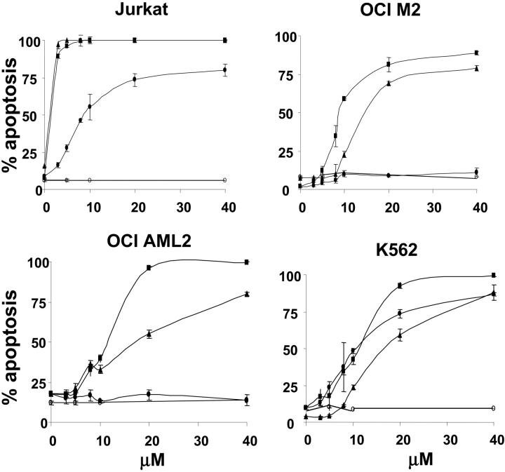
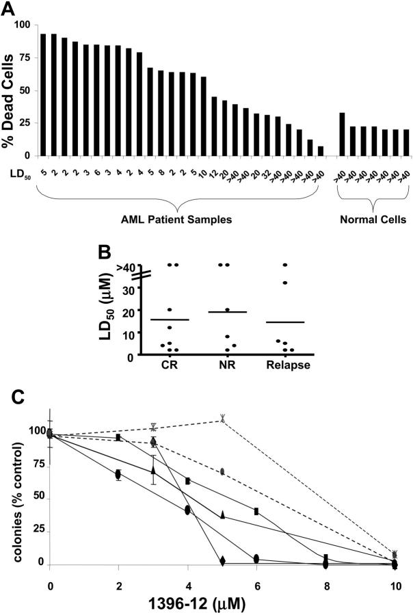
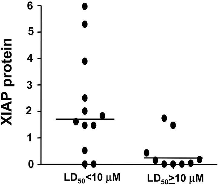
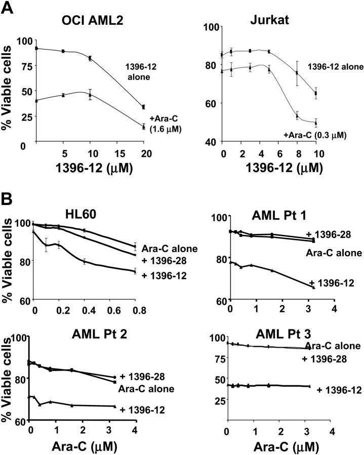
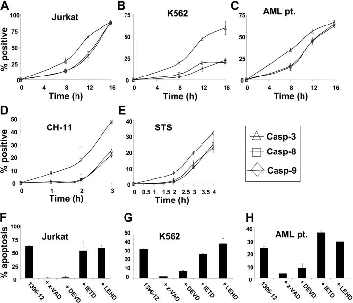
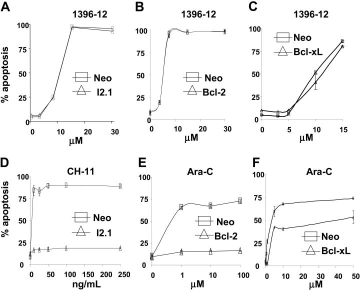
We tested the effects of small-molecule XIAP antagonists based on a polyphenylurea pharmacophore on cultured acute myelogenous leukemia (AML) cell lines and primary patient samples. X-linked inhibitor of apoptosis protein (XIAP) antagonist N-[(5R)-6-[(anilinocarbonyl)amino]-5-((anilinocarbonyl){[(2R)-1-(4-cyclohexylbutyl)pyrrolidin-2-yl]methyl}amino)hexyl]-N-methyl-N'-phenylurea (1396-12), but not a structurally related control compound, induced apoptosis of primary leukemia samples with a lethal dose (LD50) of less than 10 microM in 16 of 27 (60%) samples. In contrast, XIAP antagonist 1396-12 was not lethal to the normal hematopoietic cells in short-term cytotoxicity assays. Response of primary AML specimens to XIAP inhibitor correlated with XIAP protein levels, with higher levels of XIAP associated with sensitivity. The XIAP antagonist 1396-12 induced activation of downstream caspases 3 and 7 prior to the activation of upstream caspase 8 and caspase 9. Apoptosis induction was also independent of B-cell lymphoma protein-2 (Bcl-2) or caspase 8, indicative of a downstream effect on apoptotic pathways. Thus, polyphenylurea-based XIAP antagonsists directly induce apoptosis of leukemia cells and AML patient samples at low micromolar concentrations through a mechanism of action distinct from conventional chemotherapeutic agents.


Figures

Figure 1.
Figure 1.
XIAP antagonists induce apoptosis of leukemia cell lines. Jurkat, OCI-M2, OCI-AML 2, and K562 leukemia cells (6.5 × 10⁵/mL) were treated with increasing concentrations of the active XIAP antagonists 1396-12 (▪), 1396-22 (▴), 1396-34 (•), or the structurally related inactive control 1396-28 (○). At 24 hours after treatment, apoptosis was measured by annexin V staining (% positivity). The mean plus or minus SD of 3 independent experiments is shown.
Figure 2.
Figure 2.
XIAP inhibitor induces apoptosis in primary AML samples. (A) Primary AML blasts were isolated from peripheral blood samples obtained from patients with AML who had more than 80% blasts in the peripheral blood. As a control, mononuclear cells were isolated from samples of normal mobilized peripheral blood cells or from bone marrow. Primary blasts or normal hematopoietic mononuclear cells were treated with increasing concentrations of the XIAP inhibitor antagonist 1396-12 for 24 hours. After treatment, apoptosis was measured by annexin V surface expression. For each sample, the percentage of apoptosis after treatment with 10 μM of 1396-12 is shown with the LD50 displayed along the x-axis. (B) XIAP inhibitor is equally effective in samples from patients with chemosensitive and chemoresistant AML. The LD50 for 1396-12 is compared between samples derived from patients who achieved complete remission with induction chemotherapy (CR), had no response to induction chemotherapy (NR), or who were in relapse. The horizontal line represents the median LD50 for the group. (C) Colony formation assay. Primary AML blasts (—) or mobilized normal peripheral blood stem cells (-) were treated with increasing concentrations of the XIAP antagonist 1396-12 or buffer control for 24 hours. After treatment, cells were washed, plated in methylcellulose cultures, and counted after one week. For each sample, the mean plus or minus SD of 3 experiments expressed as a percentage of the buffer treated control is shown. Each symbol represents an individual patient.
Figure 3.
Figure 3.
Correlation of XIAP levels with in vitro response to XIAP antagonists. Protein lysates were prepared from 21 of 27 AML samples. Samples were normalized for total protein content, and analyzed by SDS-PAGE/immunoblotting using antibodies specific for XIAP and β-actin. Immunoblot data were quantified by scanning densitometry, normalized for β-actin expression, and compared with a sample of normal peripheral blood stem cells as an internal reference control. The figure shows the relative level of XIAP protein expressed as a fold increase over the reference standard for patient samples dichotomized by their response to the XIAP inhibitor (LD50 < 10 μM or LD50 ≥ 10 μM). Horizontal lines represent the median XIAP protein level for the group.
Figure 4.
Figure 4.
Studies of XIAP inhibitor 1396-12 in combination with Ara-C. (A) OCI AML2 and Jurkat leukemia cells (6.25 × 10⁵/mL) were treated with increasing concentrations of 1396-12 with (▴) or without (▪) Ara-C. Twelve hours after incubation, apoptosis was measured by annexin V staining. (B) HL60 leukemia cells (6.25 × 10⁵/mL) or primary AML patient samples (AML Pt 1-3) (× 105/mL) were treated with increasing concentrations of Ara-C alone (▪) or in combination with 8 μM (HL60) or 10 μM (AML patient samples) of the active XIAP inhibitor 1396-12 (▴) or the inactive control 1396-28 (•). Apoptosis was measured by annexin V staining 48 hours after incubation. The mean percentage plus or minus SD of viable cells is shown.
Figure 5.
Figure 5.
XIAP inhibitor 1396-12 activates effector caspases. XIAP inhibitor 1396-12 activates effector caspases as an early event: Jurkat (A), K562 (B), and primary AML (C) leukemic cells (6 × 10⁵/mL) were treated with the XIAP inhibitor 1396-12 (10 μM) for increasing periods of time. As controls, Jurkat cells were treated with CH-11 anti-Fas antibody (100 ng/mL; D) or staurosporine (STS; E) for increasing periods of time. After treatment, the mean percentage plus or minus SD of cells above control with active caspase 3 (▵), caspase 8 (□), and caspase 9 (⋄) was determined. Inhibition of effector caspases blocks XIAP inhibitor-induced apoptosis: Jurkat (F), K562 (G), and primary AML leukemic cells (H) (6 × 10⁵/mL) were treated with the XIAP inhibitor 1396-12 (10 μM) for 12 hours with or without the pan-caspase inhibitor z-VAD, the preferential effector caspase inhibitor DEVD, the preferential caspase 8 inhibitor IETD, or the preferential caspase 9 inhibitor LEHD. After treatment, apoptosis was measured by annexin V staining. The mean percentage plus or minus SD of apoptotic cells above control is shown.
Figure 6.
Figure 6.
XIAP inhibitor 1396-12 induces apoptosis independent of Bcl-2, Bcl-XL, and caspase 8. Jurkat cells lacking caspase 8 (I2.1) (A,▵), Jurkat cells overexpressing Bcl-2 (B,▵), U937 cells overexpressing Bcl-xL (C, ▵), or the corresponding vector controls (6 × 10⁵/mL; □) were treated with increasing concentrations of the XIAP inhibitor 1396-12. As controls, Jurkat cells lacking caspase 8 (D, ▵) were treated with increasing concentrations of CH-11 anti-Fas antibody (100 ng/mL). Also, Jurkat cells (E) and U937 cells (F) overexpressing Bcl-2 or Bcl-xL (▵), respectively, were treated with increasing concentrations of Ara-C. Apoptosis was measured by annexin V staining. The mean plus or minus SD of 3 independent experiments is shown.

References

    1. Green DR, Evan GI. A matter of life and death. Cancer Cell. 2002;1: 19-30. - PubMed
    1. Cryns V, Yuan Y. Proteases to die for. Genes Dev. 1999;12: 1551-1570. - PubMed
    1. Thornberry NA, Lazebnik Y. Caspases: enemies within. Science. 1998;281: 1312-1316. - PubMed
    1. Deveraux QL, Takahashi R, Salvesen GS, Reed JC. X-linked IAP is a direct inhibitor of cell death proteases. Nature. 1997;388: 300-304. - PubMed
    1. Liu Z, Sun C, Olejniczak ET, et al. Structural basis for binding of Smac/DIABLO to the XIAP BIR3 domain. Nature. 2000;408: 1004-1008. - PubMed

Publication types

Substances