. 2005 Feb 1;19(3):322-7.
doi: 10.1101/gad.1254305.

Visualization of looping involving the immunoglobulin heavy-chain locus in developing B cells

Affiliations

Visualization of looping involving the immunoglobulin heavy-chain locus in developing B cells

Camil E Sayegh et al. Genes Dev. .

Erratum in

  • Genes Dev. 2008 Jun 15;22(12):1717. Sayegh, Camil [corrected to Sayegh, Camil E]

Abstract

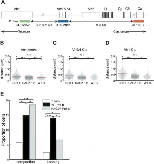
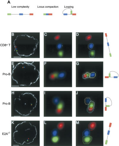
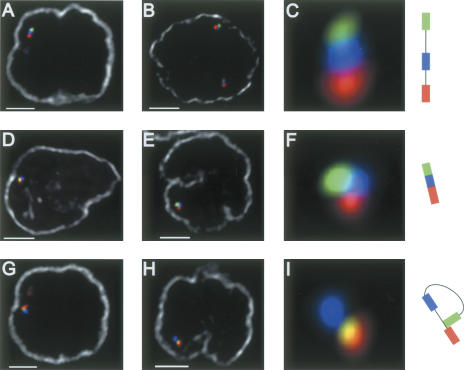
The immunoglobulin heavy-chain (IgH) locus undergoes large-scale contraction in B cells poised to undergo IgH V(D)J recombination. We considered the possibility that looping of distinct IgH V regions plays a role in promoting long-range interactions. Here, we simultaneously visualize three subregions of the IgH locus, using three-dimensional fluorescence in situ hybridization. Looping within the IgH locus was observed in both B- and T-lineage cells. However, monoallelic looping of IgH V regions into close proximity of the IgH DJ cluster was detected in developing B cells with significantly higher frequency when compared with hematopoietic progenitor or CD8+ T-lineage cells. Looping of a subset of IgH V regions, albeit at lower frequency, was also observed in RAG-deficient pro-B cells. Based on these observations, we propose that Ig loci are repositioned by a looping mechanism prior to IgH V(D)J rearrangement to facilitate the joining of Ig variable, diversity, and joining segments.


Figures

Figure 1.
Figure 1.
Compaction of distinct subregions of the IgH locus. (A) The murine IgH locus and positions of the three BAC probes are indicated (not drawn to scale). The distances separating each of the three BAC probes—CT7-526A21, CT7-34H6, and RP23-24I12—and their positions within the IgH locus were determined using the Ensembl mouse genome database. Note that the exact location of the CT7-526A21 BAC remains to be determined. The colors of the three probes are shown as follows: CT7-526A21 (green), RP23-24I12 (blue), and CT7-34H6 (red). (B-D) Scatter-plots of the distances separating the V and Cα regions in T cells, in vitro-cultured wild-type, and RAG2-deficient pro-B cells. Y-axis indicates distances separating the loci in microns. Red line indicates the average distance in each group. (★★★) Significant difference (p < 0.0001) of the averages compared with that obtained from T-lineage cells. (E) Proportion of cells (Y-axis) in which the IgH locus is compacted or in a looping configuration in T cells (white), wild-type pro-B cells (black), or RAG2-/- pro-B cells (gray). (nd) Not determined; () p < 0.05; (★★) p < 0.01.
Figure 2.
Figure 2.
Looping involving the distal IgH Vh1 and the Cα regions. (A) Schematic diagram of three possible distinct IgH configurations. (B-M) Three-dimensional configuration of the IgH locus resolved using four-color FISH. Digitally magnified pictures of IgH alleles are shown. Polygons (in one selected z-section) that were used to identify the coordinates of the center of mass for each signal are indicated (D,G,J,M). The nuclear membrane stained using lamin antibodies is shown in gray (B,E,H,K). Bar, 2 μm. (B-D) IgH locus in CD8+ T cells. (E-J) Looping involving the Vh1 and the Cα region in wild-type pro-B cells. (K-M) Configuration of the IgH locus in E2A-deficient hematopoietic progenitor cells.
Figure 3.
Figure 3.
Looping involving the IgH locus in RAG-deficient pro-B cells. Three distinct configurations of the IgH locus were detected Three-dimensional FISH in nuclei derived RAG deficient pro-B cells. (A-C) Low-complexity. (D-F) Locus compaction. (G-I) Looping. The nuclear membrane is shown in gray. Bar, 2 μm.

References

    1. Baxter J., Merkenschlager, M., and Fisher, A.G. 2002. Nuclear organisation and gene expression. Curr. Opin. Cell Biol. 14: 372-376. - PubMed
    1. Bergman Y. and Cedar, H. 2004. A stepwise epigenetic process controls immunoglobulin allelic exclusion. Nat. Rev. Immunol. 4: 753-761. - PubMed
    1. Carter D., Chakalova, L., Osborne, C.S., Dai, Y.F., and Fraser, P. 2002. Long-range chromatin regulatory interactions in vivo. Nat. Genet. 32: 623-626. - PubMed
    1. Chevillard C., Ozaki, J., Herring, C.D., and Riblet, R. 2002. A three-megabase yeast artificial chromosome contig spanning the C57BL mouse Igh locus. J. Immunol. 168: 5659-5666. - PubMed
    1. Chowdhury D. and Sen, R. 2001. Stepwise activation of the immunoglobulin μ heavy chain gene locus. EMBO J. 20: 6394-6403. - PMC - PubMed

Publication types

MeSH terms

Substances