. 2005 Feb 1;33(2):e22.
doi: 10.1093/nar/gni023.

Tetracycline-inducible gene regulation in mycobacteria

Affiliations

Tetracycline-inducible gene regulation in mycobacteria

Marian C J Blokpoel et al. Nucleic Acids Res. .

Abstract

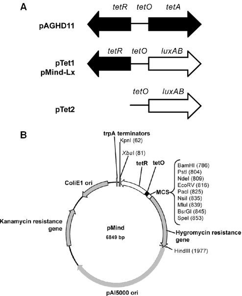
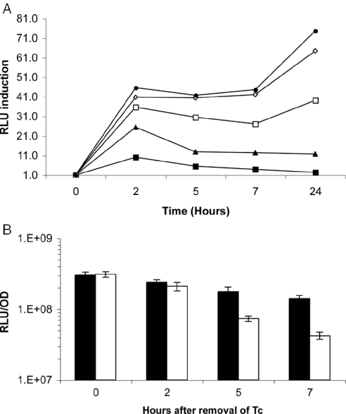
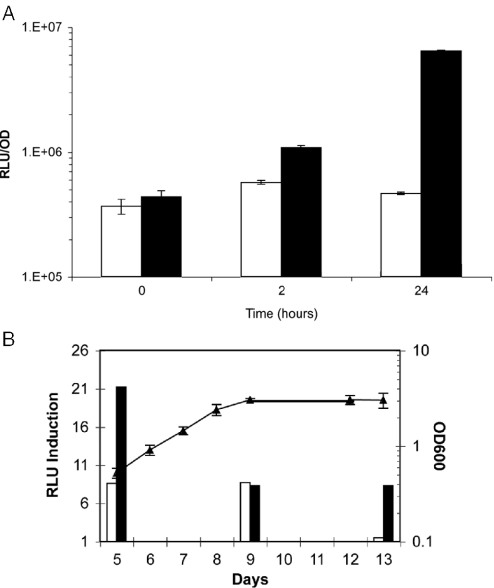
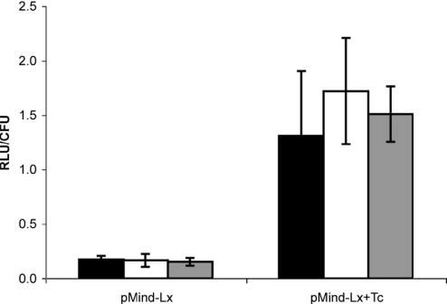
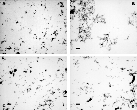
A system for the tetracycline-inducible regulation of gene expression in mycobacteria has been developed. We have sub-cloned the tetRO region from the Corynebacterium glutamicum TetZ locus into a mycobacterial shuttle plasmid, making expression of genes cloned downstream of tetRO responsive to tetracycline. Using the luxAB-encoded luciferase from Vibrio harveyi as a reporter (pMind-Lx), we observed a 40-fold increase in light output from Mycobacterium smegmatis cultures 2 h after adding 20 ng ml(-1) of tetracycline. Similarly, exposure to the drug resulted in up to 20-fold increase in relative light units from M.bovis BCG carrying the reporter construct, and a 10-fold increase for M.tuberculosis. Tetracycline induction was demonstrated in log and stationary phase cultures. To evaluate whether this system is amenable to use in vivo, J774 macrophages were infected with M.bovis BCG[pMind-Lx], treated with amikacin to kill extracellular bacteria, and then incubated with tetracycline. A 10-fold increase in light output was measured after 24 h, indicating that intracellular bacteria are accessible and responsive to exogenously added tetracycline. To test the use of the tetracycline-inducible system for conditional gene silencing, mycobacteria were transformed with a pMind construct with tetRO driving expression of antisense RNA for the ftsZ gene. Bacterial cells containing the antisense construct formed filaments after 24 h exposure to tetracycline. These results demonstrate the potential of this tetracycline-regulated system for the manipulation of mycobacterial gene expression inside and outside cells.


Figures

Figure 1
Figure 1
(A) The organization of the TetZ region from pAGHD1. pTet1 and pMind-Lx contained the tetR and tetO regions cloned in front of the luxAB reporter; tetR was omitted from pTet2. (B) A map of the pMind vector, illustrating origins of replication and selectable markers for both E.coli and mycobacteria, and the tetRO operator–repressor region upstream of a multiple cloning site.
Figure 2
Figure 2
(A) The kinetics of luciferase induction following addition of varying concentrations of Tc to M.smegmatis [pMind-Lx]. Tc was added to bacterial cultures and luminescence was measured after different time intervals. Tc was added at 1.25 ng ml−1 (filled square), 2.5 ng ml−1 (filled triangle), 5 ng ml−1 (open square), 10 ng ml−1 (open diamond) and 20 ng ml−1 (filled circle). Y-axis values are the ratio of RLU measured in the presence and absence of Tc. (B) The decrease in luminescence over time after Tc has been removed. Open bars show the loss of luminescence from induced M.smegmatis [pMind-Lx] that were washed and resuspended in fresh medium. The corresponding induced culture was washed then resuspended in its original Tc-containing medium (dark bars). Y-axis values are RLU per OD unit.
Figure 3
Figure 3
(A) The light output from M.tuberculosis [pMind-Lx] after 2 and 24 h induction with Tc. Samples were incubated with 20 ng ml−1 Tc (filled bars) and controls without Tc (open bars). Y-axis values are RLU per OD unit. (B) Luciferase induction from M.bovis BCG[pMind-Lx] at various times during growth. Replicate cultures were induced with 20 ng ml−1 Tc on day 5 (log phase growth), day 9 (early stationary phase) and day 13 (late stationary phase) and the light output measure after 2 h (open bars) and 24 h (filled bars). The ratio of RLU in the presence and absence of Tc induction is plotted on the Y-axis. The mean OD600nm for all the replicate cultures is also shown (filled triangles).
Figure 4
Figure 4
Luminescence produced by intra-phagosomal M.bovis BCG[pMind-Lx]. J774 macrophages infected with M.bovis BCG[pMind-Lx] were exposed to 100 ng ml−1 Tc and light output was measured in samples taken after 24, 48 or 72 h. Addition of Tc resulted in an ∼10-fold increase in light output from BCG[pMind] but had no effect on output from the constitutively expressed luxAB in BCG[pSHK-Lx] (data not shown). There was no significant difference in the number of viable bacteria present in drug-treated or control cultures. Y-axis values are mean RLU per CFU from triplicate wells; error bars represent SD values. Filled bars, 24 h; open bars, 48 h; grey bars, 72 h induction.
Figure 5
Figure 5
Micrographs showing the filamented morphology of M.smegmatis after induction of ftsZ antisense. (A) M.smegmatis [pMind-FtsZ-antisense] after 24 h without Tc; (B) M.smegmatis [pMind-FtsZ-antisense] after 24 h induction with 20 ng ml−1 Tc; (C) M.smegmatis without Tc; (D) M.smegmatis with 20 ng ml−1 Tc. Images taken under a 100× objective, the scale bar is 2 μm.

References

    1. Dye C., Scheele S., Dolin P., Pathania V., Raviglione R.C. Global burden of tuberculosis—estimated incidence, prevalence, and mortality by country. J. Am. Med. Assoc. 1999;282:677–686. - PubMed
    1. Wayne L.G., Sohaskey C.D. Nonreplicating persistence of Mycobacterium tuberculosis. Annu. Rev. Microbiol. 2001;55:139–163. - PubMed
    1. Smeulders M.J., Keer J., Speight R.A., Williams H.D. Adaptation of Mycobacterium smegmatis to stationary phase. J. Bacteriol. 1999;181:270–283. - PMC - PubMed
    1. Capuano S.V., III, Croix D.A., Pawar S., Zinovik A., Myers A., Lin P.L., Bissel S., Fuhrman C., Klein E., Flynn J.L. Experimental Mycobacterium tuberculosis infection of cynomolgus macaques closely resembles the various manifestations of human M.tuberculosis infection. Infect. Immun. 2003;71:5831–5844. - PMC - PubMed
    1. Flynn J.L., Capuano S.V., Croix D., Pawar S., Myers A., Zinovik A., Klein E. Non-human primates: a model for tuberculosis research. Tuberculosis (Edinb.) 2003;83:116–118. - PubMed

Publication types

MeSH terms