The yeast phosphotyrosyl phosphatase activator is part of the Tap42-phosphatase complexes
- PMID: 15689491
- PMCID: PMC1073688
- DOI: 10.1091/mbc.e04-09-0797
The yeast phosphotyrosyl phosphatase activator is part of the Tap42-phosphatase complexes
Abstract
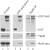
Phosphotyrosyl phosphatase activator PTPA is a type 2A phosphatase regulatory protein that possesses an ability to stimulate the phosphotyrosyl phosphatase activity of PP2A in vitro. In yeast Saccharomyces cerevisiae, PTPA is encoded by two related genes, RRD1 and RRD2, whose products are 38 and 37% identical, respectively, to the mammalian PTPA. Inactivation of either gene renders yeast cells rapamycin resistant. In this study, we investigate the mechanism underling rapamycin resistance associated with inactivation of PTPA in yeast. We show that the yeast PTPA is an integral part of the Tap42-phosphatase complexes that act downstream of the Tor proteins, the target of rapamycin. We demonstrate a specific interaction of Rrd1 with the Tap42-Sit4 complex and that of Rrd2 with the Tap42-PP2Ac complex. A small portion of PTPA also is found to be associated with the AC dimeric core of PP2A, but the amount is significantly less than that associated with the Tap42-containing complexes. In addition, our results show that the association of PTPA with Tap42-phosphatase complexes is rapamycin sensitive, and importantly, that rapamycin treatment results in release of the PTPA-phosphatase dimer as a functional phosphatase unit.
Figures


Similar articles
-
Specific interactions of PP2A and PP2A-like phosphatases with the yeast PTPA homologues, Ypa1 and Ypa2.Biochem J. 2005 Feb 15;386(Pt 1):93-102. doi: 10.1042/BJ20040887. Biochem J. 2005. PMID: 15447631 Free PMC article.
-
Functional analysis of RRD1 (YIL153w) and RRD2 (YPL152w), which encode two putative activators of the phosphotyrosyl phosphatase activity of PP2A in Saccharomyces cerevisiae.Mol Gen Genet. 2000 Jan;262(6):1081-92. doi: 10.1007/pl00008651. Mol Gen Genet. 2000. PMID: 10660069
-
Tor proteins and protein phosphatase 2A reciprocally regulate Tap42 in controlling cell growth in yeast.EMBO J. 1999 May 17;18(10):2782-92. doi: 10.1093/emboj/18.10.2782. EMBO J. 1999. PMID: 10329624 Free PMC article.
-
Protein phosphatase 2A on track for nutrient-induced signalling in yeast.Mol Microbiol. 2002 Feb;43(4):835-42. doi: 10.1046/j.1365-2958.2002.02786.x. Mol Microbiol. 2002. PMID: 11929536 Review.
-
The role of phosphatases in TOR signaling in yeast.Curr Top Microbiol Immunol. 2004;279:19-38. doi: 10.1007/978-3-642-18930-2_2. Curr Top Microbiol Immunol. 2004. PMID: 14560949 Review.
Cited by
-
The TOR complex 1 is a direct target of Rho1 GTPase.Mol Cell. 2012 Mar 30;45(6):743-53. doi: 10.1016/j.molcel.2012.01.028. Epub 2012 Mar 22. Mol Cell. 2012. PMID: 22445487 Free PMC article.
-
The yeast Tor signaling pathway is involved in G2/M transition via polo-kinase.PLoS One. 2008 May 21;3(5):e2223. doi: 10.1371/journal.pone.0002223. PLoS One. 2008. PMID: 18493323 Free PMC article.
-
The peptidyl prolyl isomerase Rrd1 regulates the elongation of RNA polymerase II during transcriptional stresses.PLoS One. 2011;6(8):e23159. doi: 10.1371/journal.pone.0023159. Epub 2011 Aug 24. PLoS One. 2011. PMID: 21887235 Free PMC article.
-
The Saccharomyces cerevisiae phosphatase activator RRD1 is required to modulate gene expression in response to rapamycin exposure.Genetics. 2006 Feb;172(2):1369-72. doi: 10.1534/genetics.105.046110. Epub 2005 Dec 1. Genetics. 2006. PMID: 16322523 Free PMC article.
-
Ser/Thr protein phosphatases in fungi: structure, regulation and function.Microb Cell. 2019 Apr 24;6(5):217-256. doi: 10.15698/mic2019.05.677. Microb Cell. 2019. PMID: 31114794 Free PMC article. Review.
References
-
- Beck, T., and Hall, M. N. (1999). The TOR signalling pathway controls nuclear localization of nutrient-regulated transcription factors. Nature 402, 689-692. - PubMed
-
- Bertram, P. G., Choi, J. H., Carvalho, J., Ai, W., Zeng, C., Chan, T. F., and Zheng, X. F. (2000). Tripartite regulation of Gln3p by TOR, Ure2p and phosphatases. J. Biol. Chem. 275, 35727-35733. - PubMed
-
- Cayla, X., Goris, J., Hermann, J., Hendrix, P., Ozon, R., and Merlevede, W. (1990). Isolation and characterization of a tyrosyl phosphatase activator from rabbit skeletal muscle and Xenopus laevis oocytes. Biochemistry 29, 658-667. - PubMed
-
- Cayla, X., Van Hoof, C., Bosch, M., Waelkens, E., Vandekerckhove, J., Peeters, B., Merlevede, W., and Goris, J. (1994). Molecular cloning, expression, and characterization of PTPA, a protein that activates the tyrosyl phosphatase activity of protein phosphatase 2A. J. Biol. Chem. 269, 15668-15675. - PubMed
Publication types
MeSH terms
Substances
LinkOut - more resources
Full Text Sources
Other Literature Sources
Molecular Biology Databases
Miscellaneous

