. 2005 Apr;16(4):1883-900.
doi: 10.1091/mbc.e04-07-0564. Epub 2005 Feb 2.

The pleckstrin homology domain proteins Slm1 and Slm2 are required for actin cytoskeleton organization in yeast and bind phosphatidylinositol-4,5-bisphosphate and TORC2

Affiliations

The pleckstrin homology domain proteins Slm1 and Slm2 are required for actin cytoskeleton organization in yeast and bind phosphatidylinositol-4,5-bisphosphate and TORC2

Maria Fadri et al. Mol Biol Cell. 2005 Apr.

Abstract

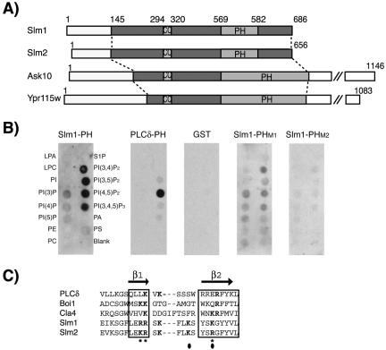
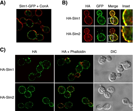
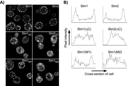
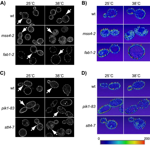
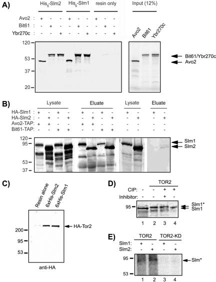
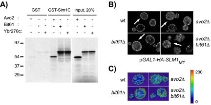
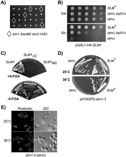
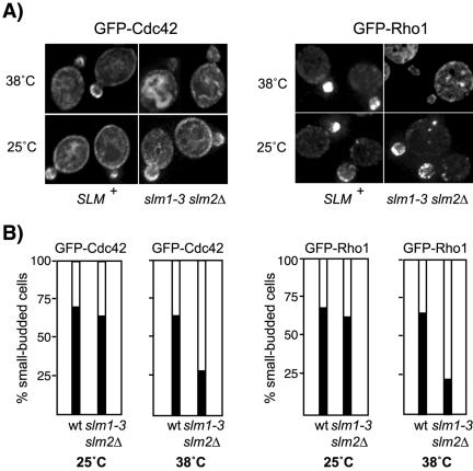
Phosphatidylinositol-4,5-bisphosphate [PtdIns(4,5)P(2)] is a key second messenger that regulates actin and membrane dynamics, as well as other cellular processes. Many of the effects of PtdIns(4,5)P(2) are mediated by binding to effector proteins that contain a pleckstrin homology (PH) domain. Here, we identify two novel effectors of PtdIns(4,5)P(2) in the budding yeast Saccharomyces cerevisiae: the PH domain containing protein Slm1 and its homolog Slm2. Slm1 and Slm2 serve redundant roles essential for cell growth and actin cytoskeleton polarization. Slm1 and Slm2 bind PtdIns(4,5)P(2) through their PH domains. In addition, Slm1 and Slm2 physically interact with Avo2 and Bit61, two components of the TORC2 signaling complex, which mediates Tor2 signaling to the actin cytoskeleton. Together, these interactions coordinately regulate Slm1 targeting to the plasma membrane. Our results thus identify two novel effectors of PtdIns(4,5)P(2) regulating cell growth and actin organization and suggest that Slm1 and Slm2 integrate inputs from the PtdIns(4,5)P(2) and TORC2 to modulate polarized actin assembly and growth.


Figures

Figure 1.
Figure 1.
Domain organization of Slm1 and Slm2 and lipid binding specificity. (A) Schematic diagram of Slm1, Slm2, Ask10, and Ypr115w structures showing the locations of PH (PH) and coiled-coil (CC) domains, respectively. Light and dark shaded boxes indicate the regions conserved among all four proteins. (B) Nitrocellulose-immobilized phospholipids (PIP Strip) were incubated with recombinant fusion proteins of GST to the C terminus of Slm1 (amino acids 441–686) containing either the wild-type PH domain (Slm1-PH) or the mutant variants PHM1, and PHM2. GST alone or a GST fusion to the PH domain of PLCδ, which specifically binds PtdIns(4,5)P2 in vitro were used as controls. Bound proteins were visualized by Western blot analysis with anti-GST antibodies. Spots contained lysophosphatidic acid (LPA), lysophosphacholine (LPC), PtdIns (PI), PtdIns(3)P, PtdIns(4)P, PtdIns(5)P, phosphatidylethanolamine (PE), phosphatidylcholine (PC), sphingosine-1-phosphate (S1P), PtdIns(3,4)P2, PtdIns(3,5)P2, PtdIns(4,5)P2, PtdIns(3,4,5)P3, phosphatic acid (PA), and phosphatidylserine (PS). (C) Sequence alignment of the β1/β2 regions of Slm1 and Slm2 PH domains with related PH domains from PLCδ, Boi1, and Cla4. Secondary structure is indicated above the sequence alignment. Bold letters indicate residues required for interaction with phosphoinositide ligands. Asterisks and closed circles indicate amino acid residues targeted for mutagenesis in SlmM1 and SlmM2 PH domain mutants, respectively.
Figure 2.
Figure 2.
Slm1 and Slm2 localize to the plasma membrane. (A) Cells containing chromosomally expressed Slm1-GFP fusion protein were mounted on glass slides and the localization of the GFP fusion protein (shown in green) was visualized by fluorescence microscopy, whereas cell walls were visualized by staining with Alexa-594–conjugated concanavalin A (ConA, shown in red). (B) HA-Slm1 and HA-Slm2 are concentrated in clusters along the plasma membrane that contain Pma1-GFP. HA-Slm1 (top, red), HA-Slm2 (bottom, red) expressed from the GAL1 promoter and chromosomally expressed Pma1-GFP (green) were visualized using antibodies directed against HA and GFP, respectively, followed by appropriate fluorescently labeled IgG. Yellow indicates signal overlap. The inset shows a magnification of the staining. (C) HA-Slm1 and HA-Slm2 do not localize to cortical actin patches. HA-Slm proteins were visualized as in B, whereas actin was stained with Alexa-594–conjugated phalloidin (shown in red). Differential interference images of the same cells are shown to the right.
Figure 3.
Figure 3.
The PH domain and phosphoinositide binding activity are necessary for plasma membrane association of Slm1 and Slm2. (A) Subcellular localization of HA-tagged wild-type and mutant variants of Slm1 and Slm2 expressed under the control of the GAL1 promoter and visualized by indirect immunofluorescence using antibodies against the HA-tag followed by Cy2-conjugated secondary antibodies. Shown are single z-sections of W303a cells expressing wild-type HA-tagged Slm1 and Slm2 (Slm1 and Slm2); C-terminally truncation mutants lacking the PH domain (Slm1ΔC and Slm2ΔC) and full-length Slm1 point mutants containing substitutions in the PH domain (Slm1M1 and Slm1M1). (B) Quantification of plasma membrane association of wild-type and mutant Slm1 and Slm2 variants. A density profile plot was generated using NIH Image 1.62. Pixel intensity was determined by a “raw average plot” of a cross section through the center of the cell from single confocal slices obtained from four to six cells.
Figure 4.
Figure 4.
Localization of Slm1 is dependent on PtdIns(4,5)P2 synthesis. The YCp-HA-SLM1 plasmid was introduced into the wild-type strains JK9-3Da (A) and W303a (C), and into isogenic strains containing mss4-2ts, fab1-2ts, pik1-83ts, and stt4-7ts temperature-sensitive mutations. Indirect immunofluorescence was performed on cells grown at 25°C or shifted to 38°C for 1.5 h by using antibodies against the HA-tag followed by Cy2-conjugated secondary antibodies. (B and D) Quantification of pixel intensity. Heightfield visualizations were performed on single z-sections of representative cells (marked with arrows) by using Amira 3.0. Maximum pixel intensity is indicated in red; minimum pixel intensity in dark blue in the color scheme.
Figure 5.
Figure 5.
Slm1 and Slm2 physically interact with Tor2 and TORC2 components. (A) In vitro-transcribed and -translated 35S-labeled Avo2, Bit61, and Ybr270c were incubated with recombinant 6His-Slm1 and 6His-Slm2 fusion proteins bound to nickel beads or with resin alone. Bound 35S-labeled proteins that remained after washing were analyzed by SDS-PAGE and autoradiography. The molecular masses of protein markers are indicated to the left. The inferred positions of Avo2, Bit61, and Ybr270c are indicated by arrows. (B) Western blot analysis of proteins associated with Tap-Avo2 and TAP-Bit61 during TAP purification experiments. Cell extracts were prepared from a strain that expressed C-terminally TAP-tagged Avo2 or Bit61 and contained HA-Slm1 or HA-Slm2 expressed under the control of the GAL1 promoter and were then subjected to sequential purifications on IgG and calmodulin affinity resins. Bound proteins were eluted with SDS-PAGE buffer, separated on 10% SDS-PAGE gels, and immunoblotted with antibodies directed against HA. The inferred positions of HA-Slm1 and HA-Slm2 are indicated by arrows. (C) HA-Tor2 associate with Slm1 and Slm2. Clarified cell lysates from a wild-type strain expressing HA-TOR2 were incubated with nickel beads lacking or containing bound 6His-Slm1 or 6His-Slm2. Bound proteins were subjected to SDS-PAGE analysis and Western blotting with HA monoclonal antibody (mAb). The inferred position of HA-Tor2 is indicated by an arrow. D) Yeast cell extracts from strain JK9–3D transformed with vector alone (lane 1) or HA-TOR2 (lanes 2–4) were incubated with alkaline phosphatase (CIP) in the presence (+) or absence (-) of phosphatase inhibitor cocktail as indicated, before SDS-PAGE analysis and Western blotting with HA mAb. The asterisk indicates the phosphorylated form. (E) Recombinant 6His-Slm1 (lanes 1 and 3) and 6His-Slm2 (lanes 2 and 4) were incubated with HA-Tor2 or HA-Tor2KD immunoprecipitated from cell lysates in the presence of [γ-32P]ATP. After SDS-PAGE, 32P-labeled proteins (denoted with an asterisk) were visualized by autoradiography.
Figure 6.
Figure 6.
Interaction with TORC2 stabilizes Slm1 association with the plasma membrane. (A) The Slm1 C terminus can mediate interaction with TORC2 components. In vitro-transcribed and -translated 35S-labeled Avo2, Bit61, and Ybr270c were incubated with resin alone or with resin containing GST fused inframe to the Slm1 C terminus (GST-Slm1C; containing amino acids 441–686 of Slm1). Bound 35S-labeled proteins that remained after washing were analyzed by SDS-PAGE and autoradiography. The molecular masses of protein markers are indicated to the left. (B) Plasmid pJK702 (GAL1-HA-SLM1) was introduced into wild-type cells (W303a), and isogenic avo2Δ, bit61Δ, ybr270cΔ single mutant, or avo2Δ bit61Δ double mutant cells. Cells were grown to early exponential phase in raffinose medium at 25°C and HA-Slm1 expression was induced by addition of galactose and further incubation for 2 h. HA-Slm1 was visualized in fixed cells as described in Figure 3. (C) Heightfield visualizations were performed on single z-sections of representative cells (marked with arrows) by using Amira 3.0. Maximum pixel intensity is indicated in red; minimum pixel intensity in dark blue in the color scheme.
Figure 7.
Figure 7.
Slm1 and Slm2 are essential for growth and actin cytoskeleton polarization. (A) Viable meiotic progeny of the SLM1/slm1::KanMX SLM2/slm2::HIS3 heterozygous diploid strain JK507 were recovered after microdissection of spores on YPD plates that were either G418-resistant (indicative of slm1::KanMX) or histidine prototroph (indicative of slm2::HIS3), but not both (marked by diamonds). B) The lethality of slm null mutants is suppressed by conditional expression of SLM1 or by disruption of INP51. Serial dilutions of cultures of wild-type (W303a) or of slm1-3 slm2Δ (JK516) and slm1-3 slm2Δ inp51Δ (JK518) mutant cells containing galactose-inducible SLM1 (pGAL1-HA-SLM1) were spotted onto rich medium containing glucose (Glc) or galactose (Gal) as carbon source. Shown are plates incubated for 3 d. (C) Only a LEU2 plasmid containing wild-type SLM1 but not the SLM1ΔC and SLM1M2 mutant variants can support growth of strain JK513 (slm1::KanMX slm2::HIS3 YCpURA3Gal1-HA-SLM2) on medium containing 5-fluoroorotic acid (+5-FOA), which counterselects the SLM2 URA3 plasmid. (D) Wild-type and slm1-3 slm2Δ mutant cells were streaked on solid SD-Ura medium and incubated at 25 or 38°C for 3 d. (E) Loss of Slm function causes actin cytoskeletal defects. Exponentially growing slm1-3 slm2Δ yeast cells were grown at 25°C or shifted to 38°C for 2 h. Cells were fixed and stained with Alexa-594-phalloidin to visualize the actin cytoskeleton.
Figure 8.
Figure 8.
Polarized localization of Cdc42 and Rho1 is perturbed in slmΔ mutant cells. (A) The distribution of GFP-Rho1 and GFP-Cdc42 in wild-type and slm1-3 slm2Δ mutant cells is shown. Exponential cultures were grown in selective medium at 25°C followed by growth at 38°C for additional 2 h. Expression of GFP-Cdc42 was induced on SD-Leu-Met medium for 1 h at 25°C before shift to nonpermissive temperature. GFP-Cdc42 and GFP-Rho1 localization was visualized by immunofluorescence in living cells mounted in 1% agarose. (B) Approximately 60 small-budded cells expressing GFP-Cdc42 and GFP-Rho1 were scored for polarized distribution of the GFP signal to the bud (black bars) or depolarized localization to the cortex or the cytoplasm (white bars).
Figure 9.
Figure 9.
Overexpression of PKC1 suppresses the growth and actin defects of slm mutants. (A) Vector alone or 2μ plasmids containing SLM1, PKC1, BCK1, MPK1, or the activated alleles BCK1-20 and MKK1-DD were transformed into strain JK513 (slm1::KanMX slm2::HIS3/ YCpURA3::GAL1-HA-SLM2). Growth of transformants was tested on SD-Ura-Leu medium containing glucose as carbon source incubated at 30°C for 3 d. (B) Overexpression of PKC1 bypasses Slm function and supports growth of strain JK512 (slm1::KanMX slm2::HIS3/ YCpURA3::GAL1-HA-SLM1) on medium containing 5-fluoroorotic acid (+FOA). (C) Actin cytoskeleton polarization in slm1-3 slm2Δ cells (strain JK515) transformed with plasmids expressing the indicated genes. Cells were grown in SD-Ura-Leu medium, shifted to the nonpermissive temperature of 38°C for 3 h, fixed, and stained for F-actin by using Alexa-594-phalloidin. (D) Quantitation of the actin polarization defect of slm1-3 slm2Δ cells transformed with the indicated plasmids from C. Small- to medium-budded cells (∼100) were scored for their actin polarization state. Cells were classified as having a polarized (gray bars), partially polarized (black bars), or depolarized (white bars) actin cytoskeleton.

Similar articles

Cited by

References

    1. Abe, M., Qadota, H., Hirata, A., and Ohya, Y. (2003). Lack of GTP-bound Rho1p in secretory vesicles of Saccharomyces cerevisiae. J. Cell Biol. 162, 85-97. - PMC - PubMed
    1. Abraham, R. T. (2002). Identification of TOR signaling complexes: more TORC for the cell growth engine. Cell 111, 9-12. - PubMed
    1. Alberts, A. S., Bouquin, N., Johnston, L. H., and Treisman, R. (1998). Analysis of RhoA-binding proteins reveals an interaction domain conserved in heterotrimeric G protein β subunits and the yeast response regulator protein Skn7. J. Biol. Chem. 273, 8616-8622. - PubMed
    1. Andrews, P. D., and Stark, M. J. (2000). Dynamic, Rho1p-dependent localization of Pkc1p to sites of polarized growth. J. Cell Sci. 113, 2685-2693. - PubMed
    1. Audhya, A., and Emr, S. D. (2002). Stt4 PI 4-kinase localizes to the plasma membrane and functions in the Pkc1-mediated MAP kinase cascade. Dev. Cell 2, 593-605. - PubMed

Publication types

MeSH terms

Substances