

Crystal structure of a predicted phosphoribosyltransferase (TT1426) from Thermus thermophilus HB8 at 2.01 A resolution
- PMID: 15689504
- PMCID: PMC2279281
- DOI: 10.1110/ps.041229405
Crystal structure of a predicted phosphoribosyltransferase (TT1426) from Thermus thermophilus HB8 at 2.01 A resolution
Abstract
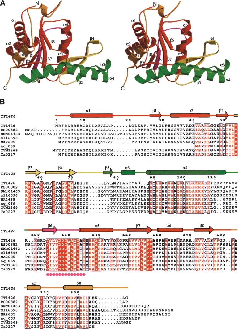
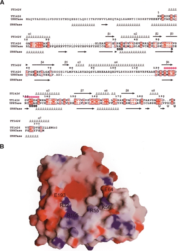
TT1426, from Thermus thermophilus HB8, is a conserved hypothetical protein with a predicted phosphoribosyltransferase (PRTase) domain, as revealed by a Pfam database search. The 2.01 A crystal structure of TT1426 has been determined by the multiwavelength anomalous dispersion (MAD) method. TT1426 comprises a core domain consisting of a central five-stranded beta sheet surrounded by four alpha-helices, and a subdomain in the C terminus. The core domain structure resembles those of the type I PRTase family proteins, although a significant structural difference exists in an inserted 43-residue region. The C-terminal subdomain corresponds to the "hood," which contains a substrate-binding site in the type I PRTases. The hood structure of TT1426 differs from those of the other type I PRTases, suggesting the possibility that TT1426 binds an unknown substrate. The structure-based sequence alignment provides clues about the amino acid residues involved in catalysis and substrate binding.
Figures
Similar articles
-
Crystal structures of possible lysine decarboxylases from Thermus thermophilus HB8.Protein Sci. 2004 Nov;13(11):3038-42. doi: 10.1110/ps.041012404. Epub 2004 Sep 30. Protein Sci. 2004. PMID: 15459330 Free PMC article.
-
Crystal structure of a purine/pyrimidine phosphoribosyltransferase-related protein from Thermus thermophilus HB8.Proteins. 2005 Nov 15;61(3):658-65. doi: 10.1002/prot.20624. Proteins. 2005. PMID: 16152602
-
ATP-induced structural change of dephosphocoenzyme A kinase from Thermus thermophilus HB8.Proteins. 2005 Jan 1;58(1):235-42. doi: 10.1002/prot.20276. Proteins. 2005. PMID: 15526298
-
Biotechnologically relevant enzymes from Thermus thermophilus.Appl Microbiol Biotechnol. 2002 Jan;58(1):1-12. doi: 10.1007/s00253-001-0843-1. Appl Microbiol Biotechnol. 2002. PMID: 11831469 Review. No abstract available.
-
Purine phosphoribosyltransferases.J Biol Chem. 2000 Jul 7;275(27):20231-4. doi: 10.1074/jbc.R000002200. J Biol Chem. 2000. PMID: 10816600 Review. No abstract available.
Cited by
-
Identification and characterization of human uracil phosphoribosyltransferase (UPRTase).J Hum Genet. 2007;52(5):415-422. doi: 10.1007/s10038-007-0129-2. Epub 2007 Mar 24. J Hum Genet. 2007. PMID: 17384901
-
Structure based annotation of Helicobacter pylori strain 26695 proteome.PLoS One. 2014 Dec 30;9(12):e115020. doi: 10.1371/journal.pone.0115020. eCollection 2014. PLoS One. 2014. PMID: 25549250 Free PMC article.
-
Local function conservation in sequence and structure space.PLoS Comput Biol. 2008 Jul 4;4(7):e1000105. doi: 10.1371/journal.pcbi.1000105. PLoS Comput Biol. 2008. PMID: 18604264 Free PMC article.
References
-
- Brünger, A.T., Adams, P.D., Clore, G.M., DeLano, W.L., Gros, P., Grosse-Kunstleve, R.W., Jiang, J.S., Kuszewski, J., Nilges, M., Pannu, N.S., et al. 1998. Crystallography and NMR system: A new software suite for macro-molecular structure determination. Acta Crystallogr. D Biol. Crystallogr. 54 905–921. - PubMed
-
- Collaborative Computational Project, Number 4. 1994. The CCP4 Suite: Programs for protein crystallography. Acta Crystallogr. D Biol. Crystallogr. 50 760–763. - PubMed
-
- Eads, J.C., Scapin, G., Xu, Y., Grubmeyer, C., and Sacchettini, J.C. 1994. The crystal structure of human hypoxanthine-guanine phosphoribosyltransferase with bound GMP. Cell 78 325–334. - PubMed
-
- Eads, J.C., Ozturk, D., Wexler, T.B., Grubmeyer, C., and Sacchettini, J.C. 1997. A new function for a common fold: The crystal structure of quinolinic acid phosphoribosyltransferase. Structure 5 47–58. - PubMed
Publication types
MeSH terms
Substances
LinkOut - more resources
Full Text Sources
Other Literature Sources
Molecular Biology Databases
