. 2005 Feb 2;25(5):1179-87.
doi: 10.1523/JNEUROSCI.1372-04.2005.

Degeneration of myelinated efferent fibers prompts mitosis in Remak Schwann cells of uninjured C-fiber afferents

Affiliations

Degeneration of myelinated efferent fibers prompts mitosis in Remak Schwann cells of uninjured C-fiber afferents

Beth B Murinson et al. J Neurosci. .

Abstract

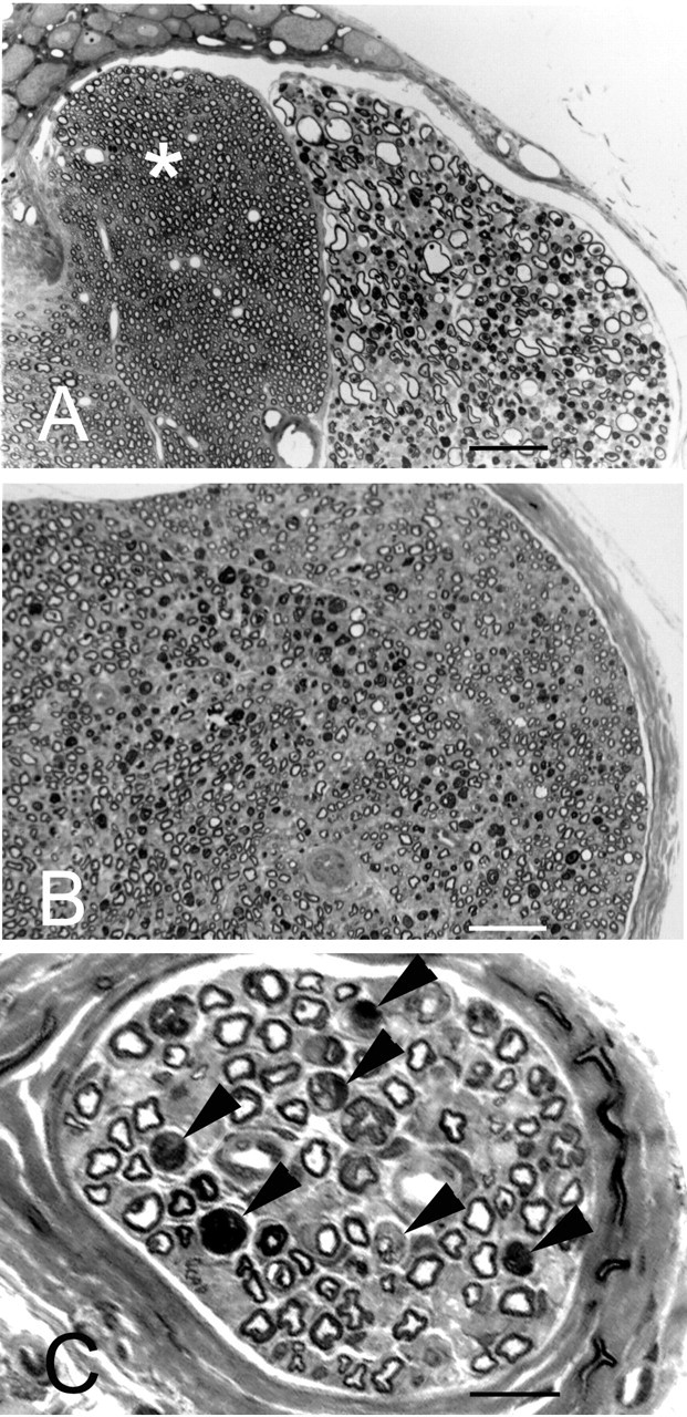
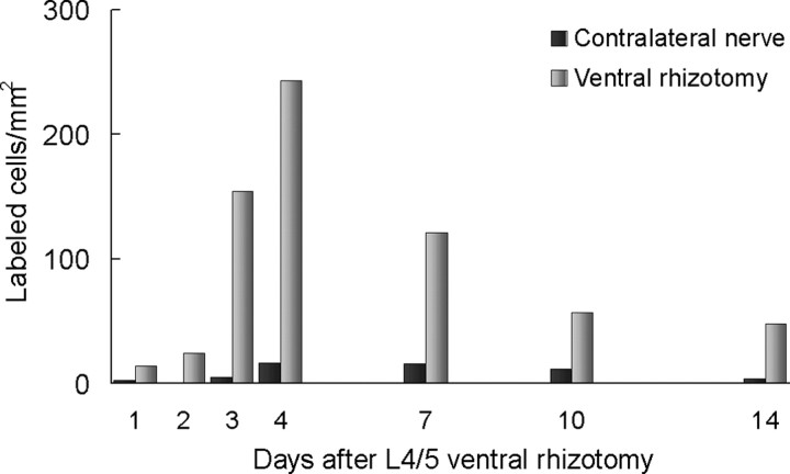
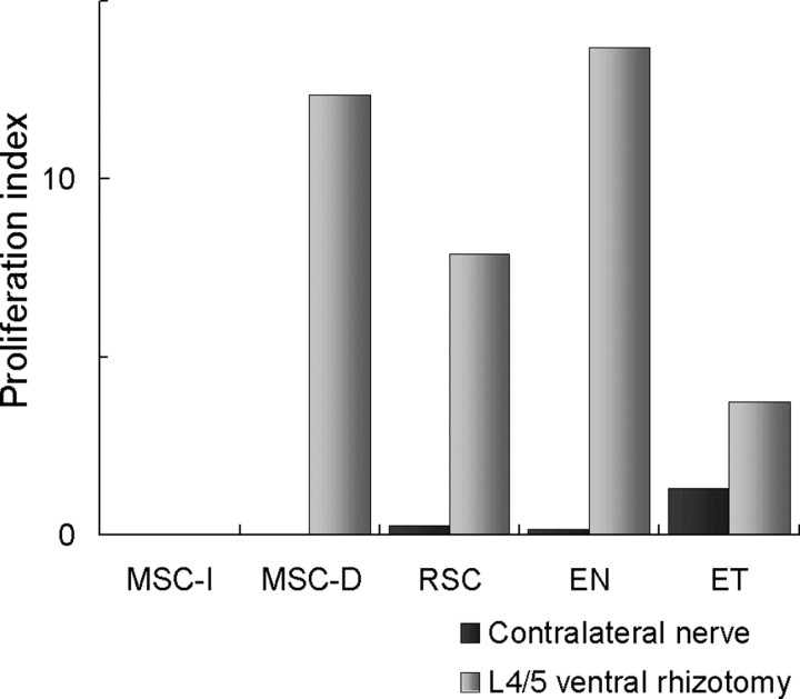
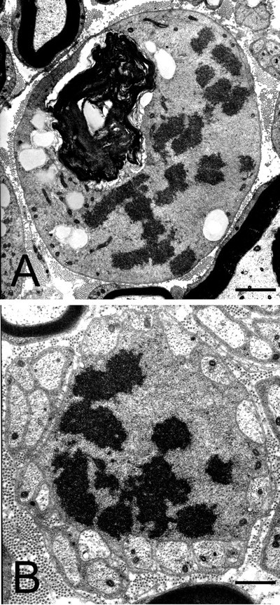
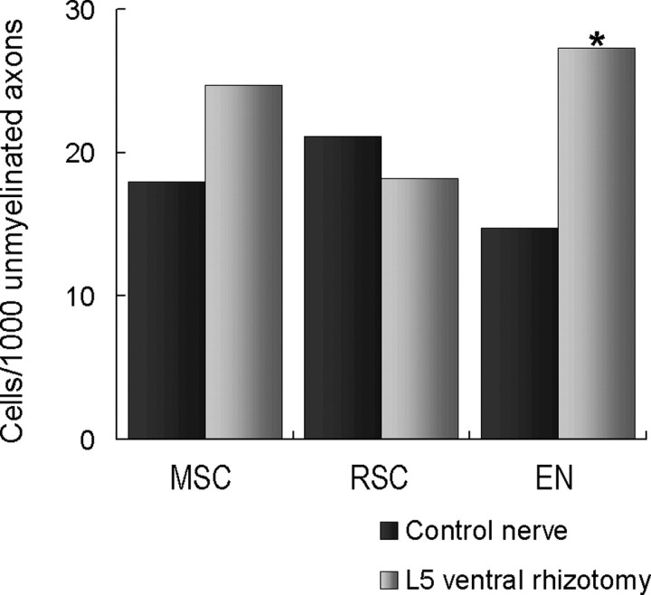
The factors inducing normally innervated Schwann cells in peripheral nerve to divide are poorly understood. Transection of the fourth and fifth lumbar ventral roots (L4/5 ventral rhizotomy) of the rat is highly selective, sparing unmyelinated axons and myelinated sensory axons; Wallerian degeneration is restricted to myelinated efferent fibers. We found that L4/5 ventral rhizotomy prompted many normally innervated nonmyelinating (Remak) Schwann cells to enter cell cycle; myelinating Schwann cells of intact (sensory) axons did not. Three days after L4/5 ventral rhizotomy, [3H]thymidine incorporation into Remak Schwann cells increased 30-fold. Schwann cells of degenerating efferents and endoneurial cells also incorporated label. Increased [3H]thymidine incorporation persisted at least 10 d after ventral rhizotomy. Despite Remak Schwann cell proliferation, the morphology of unmyelinated nerve (Remak) bundles was static. Seven days after L5 ventral rhizotomy, Remak Schwann cells in the L5-predominant lateral plantar nerve increased slightly; endoneurial cells doubled. Terminal deoxynucleotidyl transferase-mediated biotinylated UTP nick end labeling-positive nuclei increased dramatically in peripheral nerve after L5 ventral rhizotomy; many of these were macrophage nuclei. In summary, we find that the degeneration of myelinated motor axons produced signals that were mitogenic for nonmyelinating Schwann cells with intact axons but not for myelinating Schwann cells with intact axons.


Figures

Figure 1.
Figure 1.
Appearance axons in the rat lumbar outflow 7 d after ventral rhizotomy. A, Transverse section through the L5 DRG demonstrating Wallerian degeneration of axons in the ventral root; all of the axons in this root were involved. The axons in the dorsal root section (left portion of figure, asterisk) were unaffected. B, Transverse section of the sciatic nerve 7 d after L4 and L5 ventral rhizotomy demonstrating numerous myelin ovoids, increased nuclear figures, and minimal edema throughout the nerve. The degenerating fibers were scattered in islands but were in close juxtaposition to normal axons. The majority of myelinated fibers were normal in appearance. C, Lateral plantar nerve 7 d after L5 ventral rhizotomy showed decreased density of myelinated axons, some myelin ovoids, and an increased number of nuclear figures. Axons undergoing degeneration were scattered throughout the nerve (black arrowheads). The majority of myelinated axons were normal in appearance. One micrometer plastic sections were stained with toluidine blue. Scale bars: A, B, 100 μm; C, 12 μm.
Figure 2.
Figure 2.
The number of [3H]thymidine-labeled cells in the sciatic nerve at intervals after L4/5 ventral rhizotomy. The number of cells with label on the operated side is shown in light gray, and the number on the contralateral side is shown in dark gray. The number of cells incorporating [3H]thymidine increased dramatically over the first several days and did not returned to baseline 2 weeks after surgery. The values on the side contralateral to surgery were indistinguishable from control. The data are from animals labeled with schedule I (see Materials and Methods).
Figure 3.
Figure 3.
Typical electron micrographic appearance for each of the four cell types: MSC-D, the endoneurial cell, the endothelial cell, and the Remak bundle Schwann cell. The cells shown here were labeled by [3H]thymidine in rat sciatic nerve 3 d after ventral rhizotomy. In each panel, an inset image shows a light autoradiograph of the same cell that is shown in the electron micrograph, confirming [3H]thymidine uptake. A, MSC-D seen at the level of the perikaryon of a degenerating myelinated fiber. Scale bar, 1 μm. The labeling of this nucleus by [3H]thymidine is shown in the bottom right inset. Scale bar, 5 μm. This transverse section is taken through the nucleus and corresponds to a level indicated by the arrow in the teased fiber preparation in the bottom left inset. Scale bar, 3 μm. This illustrates the appearance of degenerating fibers in which the myelin debris has been displaced from the perikaryal region. B, Labeled endoneurial fibroblast. Scale bars: B, 1.5 μm; inset, 7 μm. The endoneurial cell class also included macrophages (example not shown). C, Labeled endothelial cell. Scale bars: C, 1.5 μm; inset, 7 μm. D, Labeled RSC. The nucleus that has incorporated label has a normal appearance of the Schwann cell during this premitotic phase. Scale bars: D, 1.5 μm; inset, 7 μm.
Figure 4.
Figure 4.
Proliferation index of the different cell classes in the sciatic nerve 3 d after ventral rhizotomy. There was no proliferation in MSC-I. The MSC-D and the endoneurial cells (EN) attained a high rate of [3H]thymidine incorporation, the RSC showed a substantial increase in rate of proliferation after ventral rhizotomy, and the endothelial cell population (ET) showed a minimal increase. The proliferation indices are shown for contralateral nerve (dark gray) and for post-rhizotomy (light gray).
Figure 5.
Figure 5.
Electron micrographs of mitotic Schwann cells in sciatic nerve after L4/5 ventral rhizotomy. A, Myelinating Schwann cell associated with axon undergoing Wallerian degeneration is characterized by highly condensed chromatin and the absence of a nuclear membrane. Scale bar, 3 μm. B, In a Remak Schwann cell ensheathing 14 unmyelinated axons while undergoing mitosis, nuclear contents are within nanometers of the Schwann cell membrane. Scale bar, 1.2 μm.
Figure 6.
Figure 6.
The number of cells of each type in the endoneurium of the lateral plantar nerve branch 7 d after L5 ventral rhizotomy. The columns are labeled as follows: MSC, myelinating Schwann cells; RSC, Remak (nonmyelinating) Schwann cells; EN, endoneurial cells. The data from control animals are shown as dark gray bars, and the ventral rhizotomy data are shown as light gray bars. The number of MSCs did not increase significantly. Despite evidence that ventral rhizotomy prompts vigorous proliferation among RSC, the number of these cells showed no significant increase. The numbers of endoneurial cells in the lateral plantar nerve branch doubled 1 week after L5 ventral rhizotomy.

Similar articles

Cited by

References

    1. Abercrombie M, Evans DHL, Murray LG (1959) Nuclear multiplication and cell migration in degenerating unmyelinated nerves. J Anat 93: 9-14. - PMC - PubMed
    1. Asbury AK (1967) Schwann cell proliferation in developing mouse sciatic nerve. A radioautographic study. J Cell Biol 34: 735-743. - PMC - PubMed
    1. Avellino AM, Hart D, Dailey AT, MacKinnon M, Ellegala D, Kliot M (1995) Differential macrophage responses in the peripheral and central nervous system during wallerian degeneration of axons. Exp Neurol 136: 183-198. - PubMed
    1. Baichwal RR, DeVries GH (1989) A mitogen for Schwann cells is derived from myelin basic protein. Biochem Biophys Res Commun 164: 883-888. - PubMed
    1. Baichwal RR, Bigbee JW, DeVries GH (1988) Macrophage-mediated myelin-related mitogenic factor for cultured Schwann cells. Proc Natl Acad Sci USA 85: 1701-1705. - PMC - PubMed

Publication types

MeSH terms

LinkOut - more resources