. 2005 Feb 2;25(5):1281-90.
doi: 10.1523/JNEUROSCI.4086-04.2005.

Calmodulin-dependent kinase kinase/calmodulin kinase I activity gates extracellular-regulated kinase-dependent long-term potentiation

Affiliations

Calmodulin-dependent kinase kinase/calmodulin kinase I activity gates extracellular-regulated kinase-dependent long-term potentiation

John M Schmitt et al. J Neurosci. .

Abstract

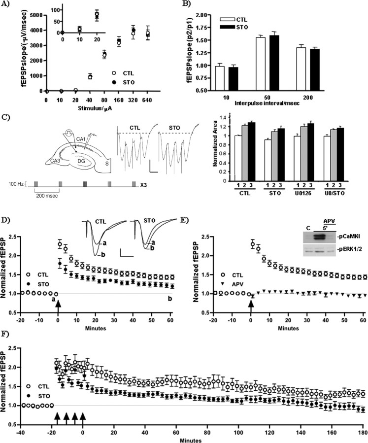
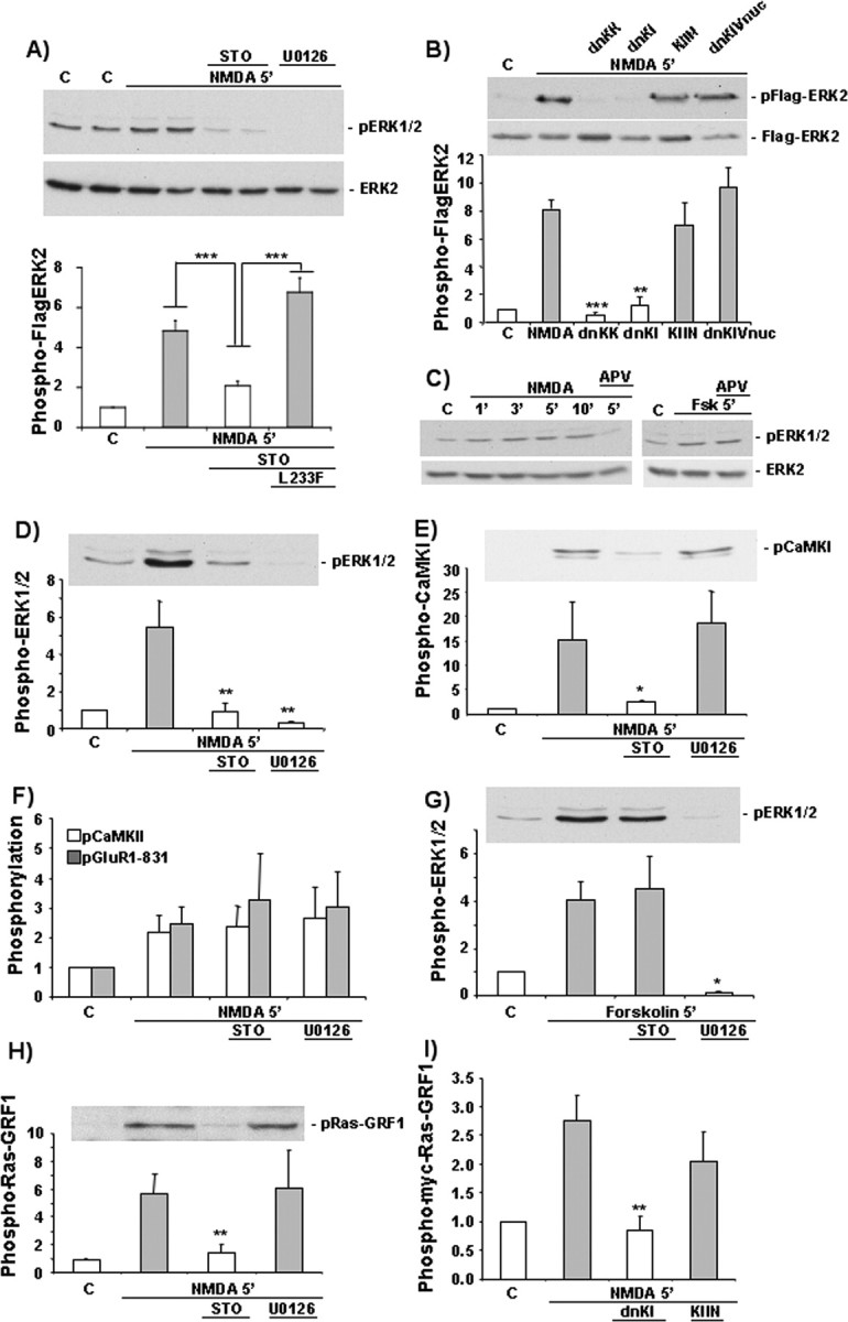
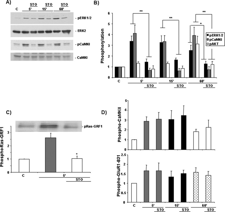
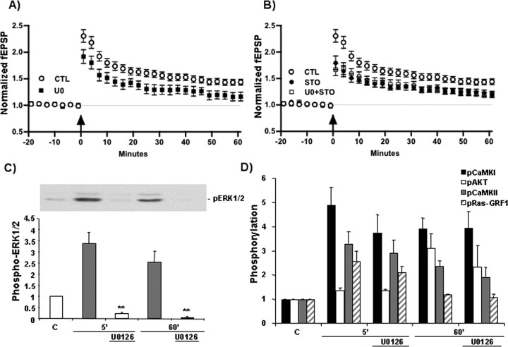
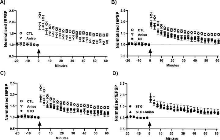
Intracellular Ca2+ and protein phosphorylation play pivotal roles in long-term potentiation (LTP), a cellular model of learning and memory. Ca2+ regulates multiple intracellular pathways, including the calmodulin-dependent kinases (CaMKs) and the ERKs (extracellular signal-regulated kinases), both of which are required for LTP. However, the mechanism by which Ca2+ activates ERK during LTP remains unknown. Here, we describe a requirement for the CaMK-kinase (CaMKK) pathway upstream of ERK in LTP induction. Both the pharmacological inhibitor of CaMKK, STO-609, and dominant-negative CaMKI (dnCaMKI), a downstream target of CaMKK, blocked neuronal NMDA receptor-dependent ERK activation. In contrast, an inhibitor of CaMKII and nuclear-localized dnCaMKIV had no effect on ERK activation. NMDA receptor-dependent LTP induction robustly activated CaMKI, the Ca2+-stimulated Ras activator Ras-GRF1 (Ras-guanyl-nucleotide releasing factor), and ERK. STO-609 blocked the activation of all three enzymes during LTP without affecting basal synaptic transmission, activation of CaMKII, or cAMP-dependent activation of ERK. LTP induction itself was suppressed 50% by STO-609 in a manner identical to the ERK inhibitor U0126: either inhibitor occluded the effect of the other, suggesting they are part of the same signaling pathway in LTP induction. STO-609 also suppressed regulatory phosphorylation of two downstream ERK targets during LTP, the general translation factors eIF4E (eukaryotic initiation factor 4) and its binding protein 4E-BP1 (eukaryotic initiation factor 4E-binding protein 1). These data indicate an essential role for CaMKK and CaMKI to link NMDA receptor-mediated Ca2+ elevation with ERK-dependent LTP.


Figures

Figure 2.
Figure 2.
Inhibition of CaMKK markedly attenuates NMDA receptor-dependent LTP. A, STO-609 does not affect basal synaptic transmission. Input-output relationship for Schaffer collateral stimulation and fEPSP initial slopes recorded from area CA1 of mouse hippocampal slices preincubated without or with 5 μm STO-609 (STO) for 30 min. Inset, 0-20 μA; n = 8. B, Paired-pulse facilitation is normal during STO-609 treatment. Paired-pulse facilitation at 10, 50, and 200 ms interpulse intervals. Values presented are ratios of fEPSP initial slopes (pulse 2/pulse 1). n = 8. C, Left, Schematic drawing of electrode placements during recording and E-LTP induction paradigm (see Materials and Methods). DG, Dentate gyrus; S, stratum radiatum. Middle, Representative first burst responses from mock-treated (control) and treated (STO) slices. Calibration: 0.5 mV, 10 ms. Right, Integrated dendritic theta-burst responses of control slices and treated slices: n = 24, control; n = 11, STO-609; n = 10, U0126; n = 10, U0126 plus STO-609 UO/STO. The theta-burst responses were calculated by integrating the fEPSP response during stimulation of naive slices with our E-LTP-generating protocol. D, E, Inhibition of CaMKK partially inhibits NMDA receptor-dependent E-LTP. After 20 min of stable baseline recording (1 test pulse per minute min), slices were treated for 30 min without or with 5 μm STO-609 for 30 min (D) or with 50 μm APV for 20 min (E) before and 5 min after theta-burst stimulation [4 pulses per burst (100 Hz), 5 bursts pertrain (5Hz), 3 trains (20s apart)]. STO-609 had no significant effect on baseline fEPSP amplitude or initial slope kinetics. Control slices (LTP, 144 ± 5.4%; n = 24) exhibited approximately two fold greater LTP than STO-treated slices (LTP, 120 ± 5.9%; n = 11) at 60 min, whereas APV treatment (n = 8) completely blocked LTP. D, Inset, Average of 10 responses, 1-10 min before (a) and 50-60 min after (b) LTP induction. E, Inset, Western blots from microdissected region CA1 showing pCaMKI and pERK 5 min after mock stimulation (control) or theta-burst stimulation without or with APV treatment. F, Inhibition of CaMKK blocks L-LTP. Slices were treated without or with STO-609 as in D before stimulation with recurrent theta-burst patterned activity [4 pulses per burst (100 Hz), 5 bursts per train (5 Hz), 6 trains (20 s apart), 4 epochs (5 min apart)] to generate late-phase LTP. Control, n = 6; STO-609, n = 6. CTL, Control.
Figure 1.
Figure 1.
NMDA activation of ERK and Ras-GRF1 in hippocampus requires CaMKK and CaMKI. Primary cultures of rat hippocampal neurons (A, B, I, 6 DIV) or acute mouse hippocampal slices (C-H, 8-12 weeks old) were preincubated with the indicated pharmacological reagents [STO-609 (STO), 5 μm, 60 min; U0126, 10 μm, 20 min; APV, 50 μm, 20 min] before stimulation by NMDA/glycine (25 and 1 μm, respectively) or forskolin (Fsk; 50 μm) for 5 min. A, B, I, Cultured rat hippocampal neurons (5 DIV) were cotransfected with Flag-ERK2 and either CaMKKL233F (L233F, a STO-609-insensitive mutant; A, bottom graph) or myc-Ras-GRF1 plus control vector pcDNA3 or the indicated dnCaMKs or CaMKIIN as indicated. Cells were stimulated with NMDA, and Flag-ERK2 or myc-Ras-GRF1 phosphorylation was determined (see Materials and Methods). In all experiments, the activation states of the indicated proteins were determined by Western blots using phospho-specific antibodies (see Materials and Methods). The ratio of the phospho-protein to total amount of that same protein was set equal to 1 for the control, and relative values for treatments are shown as fold stimulation over basal. Means ± SD; n = 6 (A), n = 3 (B), n = 5 (D-H), or n = 6 (I). *p ≤ 0.05; **p ≤ 0.01; ***p ≤ 0.001. C, Control.
Figure 3.
Figure 3.
LTP-activation of CaMKI, Ras-GRF1, and ERK requires CaMKK. Mouse hippocampal slices were preincubated without or with STO-609 (STO; 5 μm, 30 min) and subjected to theta-burst stimulation as in Figure 2 D. The activation states, assessed by phospho-specific antibodies (see Fig. 1), of ERK1/2, CaMKI, and AKT (A, B); Ras-GRF1 (C); and CaMKII and the GluR1 subunit of the AMPA-type glutamate receptor (a CaMKII substrate) (D) were determined at the indicated times. Mean ± SE; n = 6. *p ≤ 0.05; **p ≤ 0.01. C, Control.
Figure 4.
Figure 4.
CaMKK and ERK mediate E-LTP via a common mechanism. A, B, Mouse hippocampal slices were treated without or with the MEK inhibitor U0126 (UO; 10 μm) and/or the CaMKK inhibitor STO-609 (STO; 5 μm) by bath application as in Figure 2 D for 30 min before and 5 min after E-LTP induction. Control (CTL), n = 24; U0126, n = 10; STO-609, n = 11; U0126 plus STO-609, n = 10. C, D, At various times after E-LTP induction (as in Fig. 2 D), the activation status of ERK (C) and CaMKI, AKT, CaMKII, and Ras-GRF1 (D) was determined at the indicated times. Mean ± SE; n = 5. **p ≤ 0.01.
Figure 5.
Figure 5.
Inhibitors of CaMKK and MEK suppress LTP by a translation-dependent pathway. A, Mouse hippocampal slices were treated by bath application with the translation inhibitor anisomycin (Aniso; 40 μm) 30 min before and for the duration of recording after E-LTP (as in Fig. 2 D). n = 8. Note that the magnitude and kinetics of anisomycin suppression of LTP are identical to those of U0126 (UO; B) and STO-609 (STO; C) and are occluded by STO-609 (D). CTL, Control.
Figure 6.
Figure 6.
Translation factor activation in E-LTP requires CaMKK and ERK. Mouse hippocampal slices were preincubated with the indicated pharmacological reagents (see Fig. 1) and then subjected to E-LTP induction (as in Fig. 2 D). The activation state of translation factors eIF4E and its inhibitory binding protein 4E-BP1 were determined using phospho-specific antibodies against their respective activation sites. Mean ± SE; n = 6 (B) or n = 5 (D). STO, STO-609; U0, U0126; C, Control. *p ≤ 0.05; **p ≤ 0.01.

References

    1. Agell N, Bachs O, Rocamora N, Villalonga P (2002) Modulation of the Ras/Raf/MEK/ERK pathway by Ca2+ and calmodulin. Cell Signal 14: 649-654. - PubMed
    1. Arozarena I, Matallanas D, Berciano MT, Sanz-Moreno V, Calvo F, Munoz MT, Egea G, Lafarga M, Crespo P (2004) Activation of H-Ras in the endoplasmic reticulum by the RasGRF family guanine nucleotide exchange factors. Mol Cell Biol 24: 1516-1530. - PMC - PubMed
    1. Asaki C, Usuda N, Nakazawa A, Kametani K, Suzuki T (2003) Localization of translational components at the ultramicroscopic level at postsynaptic sites of the rat brain. Brain Res 972: 168-176. - PubMed
    1. Baouz S, Jacquet E, Accorsi K, Hountondji C, Balestrini M, Zippel R, Sturani E, Parmeggiani A (2001) Sites of phosphorylation by protein kinase A in CDC25Mm/GRF1, a guanine nucleotide exchange factor for Ras. J Biol Chem 276: 1742-1749. - PubMed
    1. Barria A, Muller D, Derkach V, Griffith LC, Soderling TR (1997) Regulatory phosphorylation of AMPA-type glutamate receptors by CaM-KII during long-term potentiation. Science 276: 2042-2045. - PubMed

Publication types

MeSH terms

Substances