Review
. 2005 Feb;6(2):128-33.
doi: 10.1038/sj.embor.7400337.

The dynamic organization of gene-regulatory machinery in nuclear microenvironments

Affiliations
Review

The dynamic organization of gene-regulatory machinery in nuclear microenvironments

Sayyed K Zaidi et al. EMBO Rep. 2005 Feb.

Abstract

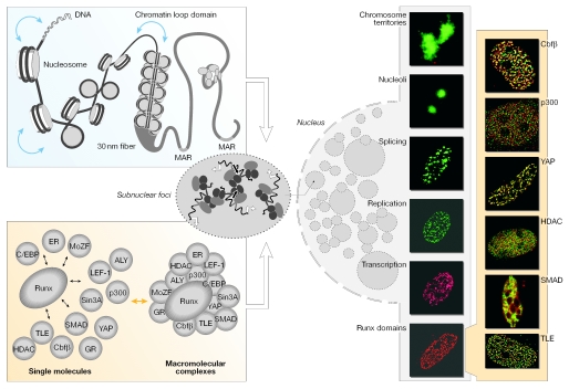
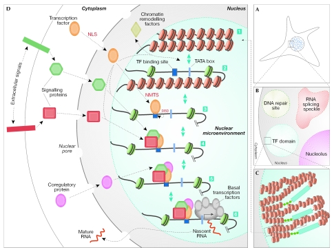
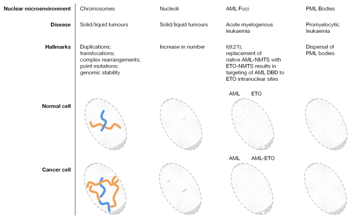
Nuclear components are functionally linked with the dynamic temporal and spatial compartmentalization, sorting and integration of regulatory information to facilitate its selective use. For example, the subnuclear targeting of transcription factors to punctate sites in the interphase nucleus mechanistically couples chromatin remodelling and the execution of signalling cascades that mediate gene expression with the combinatorial assembly of the regulatory machinery for biological control. In addition, a mitotic cycle of selective partitioning and sequential restoration of the transcriptional machinery provides a basis for the reassembly of regulatory complexes to render progeny cells competent for phenotypic gene expression. When this intranuclear targeting and localization of regulatory proteins is compromised, diseases, such as cancer, can occur. A detailed understanding of this process will provide further options for diagnosis and treatment.


Figures

Figure 1
Figure 1
Levels of nuclear organization. The linear placement of DNA-regulatory elements in gene promoters constitutes the primary level of nuclear organization. The distance between these regulatory sites is intricately regulated by the packaging of DNA into nucleosomes and higher order chromatin structures (left, upper panel). Scaffolding nuclear proteins, such as RUNX, provide structural platforms for the assembly of multiprotein supercomplexes to facilitate the combinatorial control of gene expression (left, bottom panel). Genes and macromolecular regulatory complexes together give rise to dynamic nuclear microenvironments in the nucleus. RUNX bodies are nuclear microenvironments that contain various co-regulatory proteins that are involved in gene activation, as well as repression, chromatin remodelling and cellular signalling (immunofluorescence images on the right, shaded yellow). RUNX was visualized using the Alexa 488 secondary antibody in all images and the proteins were detected using Alexa 568 fluorochrome-conjugated secondary antibodies, as indicated.
Figure 2
Figure 2
Biogenesis and regulatory activities of nuclear microenvironments that support biological control. The multistep organization and assembly of nuclear microenvironments that integrate extracellular and intracellular regulatory signals to establish transcriptional competency is shown. (A) The mammalian nucleus is functionally compartmentalized into punctate subnuclear domains (blue circles). (B) An enlarged area of the mammalian nucleus shows four nuclear microenvironments with distinct functions: the nucleolus (purple circle) is the site of ribosomal RNA synthesis; RNA-splicing speckles (shown in red), such as SC35, are involved in RNA splicing/processing; various proteins accumulate at sites of DNA damage to organize distinct nuclear domains for DNA repair (green circle); and transcription factors are present in distinct foci in the nucleus, the subnuclear placement of which often correlates with gene regulation (light-blue circle). (C) A transcription domain can contain several target genes (shown here in repressed or closed chromatin conformations) for a transcription factor to amplify the representation of the regulatory machinery, thereby facilitating the combinatorial control of transcriptional regulation. (D) This composite schematic for the control of gene expression in the context of nuclear architecture incorporates various regulatory parameters that are operative in vivo, although not necessarily in a specific temporal sequence. (1) Transcriptional repression is mediated by nucleosomes that restrict the access of promoter elements to regulatory proteins. Inactive genes show the closed chromatin conformation (red circles). (2) To accommodate cellular response to specific extracellular stimuli, chromatin modifications, which are mediated by macromolecular chromatin-remodelling complexes, render the open conformation to the gene-promoter region; this results in the accessibility of promoter domains to the tissue-restricted, as well as the basal, transcription machinery. (3) Transcription factors that are synthesized in the cytoplasm translocate into the nucleus through a nuclear-localization signal (NLS), are targeted to the subnuclear domains by the intranuclear-targeting signal (NMTS) and bind DNA through the DNA-binding domain (DBD). (4) Various physiological cues, with positive (green hexagon) or negative (red square) regulatory potential, are transmitted to the nucleus through signalling proteins; these proteins interact with transcription factors and use their subnuclear targeting signals, as well as DBDs, to converge at the target genes. (5) Other regulatory proteins (purple circle) also converge at independent, or similar, promoter regions of the target genes, which results in the combinatorial control of gene expression. (6) Finally, the basal transcription machinery is recruited to the promoter, which leads to the activation of genes and mRNA synthesis (shown as nascent RNA). The nascent RNA is then exported from the nucleus, after various steps of RNA splicing and processing that are carried out in RNA-splicing speckles, as mature RNA.
Figure 3
Figure 3
The catastrophic consequences of perturbations in nuclear microenvironments. Examples of nuclear microenvironments that undergo alterations under pathological conditions are shown. Chromosomes undergo extensive changes, including translocations, duplications and mutations, in a broad range of cancers. Similarly, the number of nucleoli, which are the sites of ribosomal gene expression, is increased in tumours. The (8;21) translocation in acute myeloid leukaemia (AML) results in the misrouting of RUNX1 to AML–eight twenty one (ETO) subnuclear sites (yellow circles). Similarly, promyelocytic leukaemia (PML) bodies increase in number and decrease in size in PML patients.

References

    1. Barseguian K, Lutterbach B, Hiebert SW, Nickerson J, Lian JB, Stein JL, van Wijnen AJ, Stein GS (2002) Multiple subnuclear targeting signals of the leukemia-related AML1/ETO and ETO repressor proteins. Proc Natl Acad Sci USA 99: 15434–15439 - PMC - PubMed
    1. Baserga R, Peruzzi F, Reiss K (2003) The IGF-1 receptor in cancer biology. Int J Cancer 107: 873–877 - PubMed
    1. Berube NG, Jagla M, Smeenk C, De Repentigny Y, Kothary R, Picketts DJ (2002) Neurodevelopmental defects resulting from ATRX overexpression in transgenic mice. Hum Mol Genet 11: 253–261 - PubMed
    1. Brown KE, Baxter J, Graf D, Merkenschlager M, Fisher AG (1999) Dynamic repositioning of genes in the nucleus of lymphocytes preparing for cell division. Mol Cell 3: 207–217 - PubMed
    1. Choi J-Y et al. (2001) Subnuclear targeting of Runx/Cbfa/AML factors is essential for tissue-specific differentiation during embryonic development. Proc Natl Acad Sci USA 98: 8650–8655 - PMC - PubMed

Publication types

MeSH terms