. 2005 Feb;115(2):291-301.
doi: 10.1172/JCI22681.

Adipocytes from Munc18c-null mice show increased sensitivity to insulin-stimulated GLUT4 externalization

Affiliations

Adipocytes from Munc18c-null mice show increased sensitivity to insulin-stimulated GLUT4 externalization

Hajime Kanda et al. J Clin Invest. 2005 Feb.

Abstract

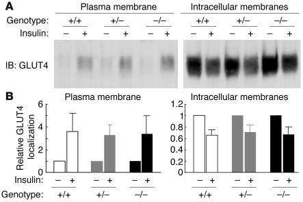
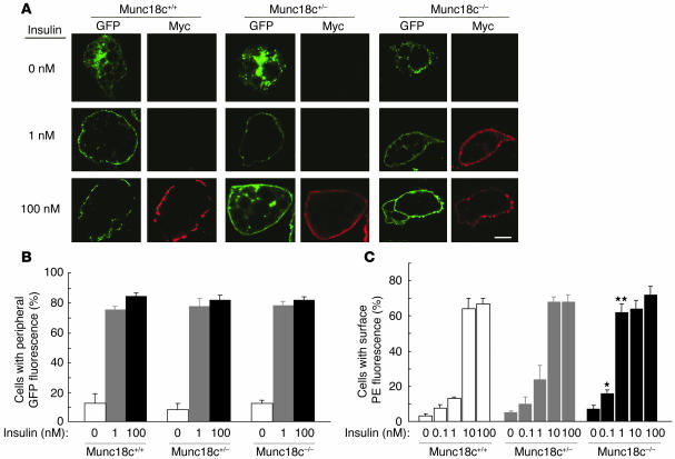
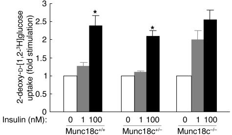
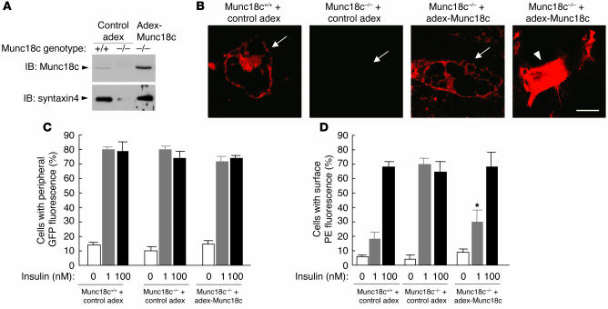
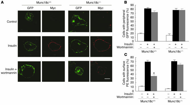
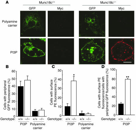
Insulin-stimulated glucose uptake in adipocytes is mediated by translocation of vesicles containing the glucose transporter GLUT4 from intracellular storage sites to the cell periphery and the subsequent fusion of these vesicles with the plasma membrane, resulting in the externalization of GLUT4. Fusion of the GLUT4-containing vesicles with the plasma membrane is mediated by a soluble N-ethylmaleimide-sensitive factor attachment protein receptor (SNARE) complex consisting of vesicle-associated membrane protein 2 (VAMP2), 23-kDa synaptosomal-associated protein (SNAP23), and syntaxin4. We have now generated mouse embryos deficient in the syntaxin4 binding protein Munc18c and show that the insulin-induced appearance of GLUT4 at the cell surface is enhanced in adipocytes derived from these Munc18c-/- mice compared with that in Munc18c+/+ cells. Wortmannin, an inhibitor of PI3K, inhibited insulin-stimulated GLUT4 externalization, without affecting GLUT4 translocation to the cell periphery, in Munc18c+/+ adipocytes, but it did not affect GLUT4 externalization in Munc18c-/- cells. Phosphatidylinositol 3-phosphate, which induced GLUT4 translocation to the cell periphery without externalization in Munc18c+/+ cells, elicited GLUT4 externalization in Munc18c-/- cells. These findings demonstrate that Munc18c inhibits insulin-stimulated externalization of GLUT4 in a wortmannin-sensitive manner, and they suggest that disruption of the interaction between syntaxin4 and Munc18c in adipocytes might result in enhancement of insulin-stimulated GLUT4 externalization.


Figures

Figure 1
Figure 1
Targeted deletion of Munc18c in mice. The wild-type Munc18c allele, the targeting vector, and the targeted allele after homologous recombination are shown. Probe A is a DNA fragment used for Southern blot analysis of BamHI-digested genomic DNA from ES cells; the wild-type and mutant alleles give rise to 9.0- and 6.4-kb hybridizing fragments, respectively.
Figure 2
Figure 2
Reduced size and disorganized brain structure of Munc18c–/– mice. (A) Gross morphology of Munc18c+/+, Munc18c+/–, and Munc18c–/– embryos at 13.5 dpc. (B) Gross morphology of Munc18c+/+, Munc18c+/–, and Munc18c–/– newborn mice. (C) Coronal sections of the brain of newborn Munc18c+/+ and Munc18c−/− mice. The sections were stained with Nissl solution. CP, cortical plate; IZ, intermediate zone; SVZ, subventricular zone. Scale bar: 200 μm.
Figure 3
Figure 3
Lack of effect of Munc18c on adipocyte differentiation in MEFs. (A) Adipocyte differentiation was induced in MEFs isolated from Munc18c+/+, Munc18c+/−, or Munc18c−/− embryos at 13.5 dpc. Twelve days after the onset of induction, adipocytes were fixed, stained with oil red O, and examined both macroscopically (Macro) and microscopically (Micro). Scale bars: 10 mm (left panels) and 50 μm (right panels). (B) Intracellular triglyceride (TG) content was quantified as described in Methods. The amount of TG content in adipocytes of each genotype was normalized to protein concentration. Data are mean ± SE of values from 4 independent experiments and are expressed relative to the value for Munc18c+/+.
Figure 4
Figure 4
Reduced abundance of syntaxin4 in Munc18c−/− adipocytes. (A) Northern blot analysis of mRNAs for SNARE proteins. Total RNA extracted from Munc18c+/+, Munc18c+/–, or Munc18c–/– adipocytes was analyzed for hybridization with probes specific for Munc18c, syntaxin4, SNAP23, VAMP2, or Munc18b mRNAs. The region of the ethidium bromide–stained gel containing 28S ribosomal RNA (rRNA) is also shown. (B) Immunoblot analysis of SNARE proteins. Lysates prepared from adipocytes of each genotype were subjected to immunoblot analysis with antibodies specific for the indicated proteins.
Figure 5
Figure 5
Increased amount of Munc18c-free syntaxin4 in Munc18c–/– adipocytes. (A) Immunoprecipitation of Munc18c-free syntaxin4 by the mAb A6C. COS-7 cells were transfected with expression plasmids for full-length rat synatxin4 and mouse Munc18c, after which cell lysates were subjected to immunoprecipitation with the A6C antibody (lane 2) or with polyclonal antibodies (pAb) to syntaxin4 (lane 3). The resulting precipitates, as well as the original cell lysate (lane 1), were subjected to immunoblot analysis with polyclonal antibodies to syntaxin4 (lower panel) and with polyclonal antibodies to Munc18c (upper panel). Munc18c was not associated with syntaxin4 immunoprecipitated by A6C (lane 2) but was present in the immunoprecipitate prepared with the polyclonal antibodies to syntaxin4 (lane 3). (B and C) Quantitation of Munc18c-free syntaxin4 in adipocytes. Lysates of Munc18c+/+, Munc18c+/–, or Munc18c–/– adipocytes (B) as well as immunoprecipitates prepared from such lysates with mAb A6C (C) were subjected to immunoblot analysis with polyclonal antibodies to syntaxin4 or to Munc18c. The bands corresponding to total syntaxin4 (B) and to Munc18c-free syntaxin4 (C) were quantitated and expressed relative to the corresponding values for Munc18c+/+ cells. Data are mean ± SE of values from 4 independent experiments. *P < 0.05, **P < 0.001 versus Munc18c+/+ cells (Student’s t test).
Figure 6
Figure 6
Lack of effect of Munc18c deficiency on proximal insulin signaling in adipocytes. Munc18c+/+, Munc18c+/–, or Munc18c–/– adipocytes were deprived of serum for 2 hours and then stimulated with insulin (1 or 100 nM) for 5 minutes. For detection of tyrosine-phosphorylated forms of IRS (pY-IRS) and the β subunit of the insulin receptor (pY-IRβ), cell lysates were subjected to immunoprecipitation and subsequent immunoblot analysis with mAb PY20 to phosphotyrosine (upper panel). For detection of serine-phosphorylated Akt, cell lysates were subjected to immunoblot analysis with antibodies specific for Akt phosphorylated on serine 473 (lower panel).
Figure 7
Figure 7
Insulin-stimulated translocation of endogenous GLUT4 to the plasma membrane in MEF-derived adipocytes. (A) Munc18c+/+, Munc18c+/–, or Munc18c–/– adipocytes were deprived of serum for 2 hours and then stimulated with 100 nM insulin for 20 minutes. Cell homogenates were subjected to subcellular fractionation, and plasma membrane and intracellular membrane fractions were subjected to immunoblot analysis with antibodies to GLUT4. (B) Immunoblot band intensity of GLUT4 was quantitated using NIH Image software and was expressed relative to the abundance of GLUT4 without insulin in cells of each genotypes. Data are mean ± SE of values from 3 or 4 independent experiments.
Figure 8
Figure 8
Enhancement of insulin-stimulated GLUT4 externalization, but not of GLUT4 translocation to the cell periphery, in Munc18c−/− adipocytes. (A) Munc18c+/+, Munc18c+/–, or Munc18c–/– adipocytes were infected with a retroviral vector encoding a GFP fusion protein of Myc epitope–tagged GLUT4 (GLUT4-myc7-GFP). The cells were stimulated with the indicated concentrations of insulin for 20 minutes, fixed, and subjected to indirect immunofluorescence staining with antibodies to the Myc tag and PE-conjugated secondary antibodies in order to detect externalized GLUT4 (red). GLUT4 translocation to the cell periphery was detected by GFP fluorescence (green) on analysis of the cells by confocal microscopy. Scale bar: 10 μm. (B) Movement of GLUT4 to the cell periphery in experiments similar to that shown in A was quantitated by determination of the percentage of GFP-positive cells that manifested GFP fluorescence at the cell periphery. Data are mean ± SE of values from 3 separate experiments, with 1,000 GFP-positive cells being examined in each experiment. (C) GLUT4 externalization in experiments similar to that shown in A was quantitated by determination of the percentage of GFP-positive cells that exhibited PE fluorescence at the cell surface. Data are mean ± SE of values from 4–6 independent experiments, with 1,000 GFP-positive cells being examined in each experiment. *P < 0.05, **P < 0.001 versus the corresponding values for Munc18c+/+ cells (Student’s t test).
Figure 9
Figure 9
Enhancement of insulin-stimulated glucose transport in Munc18c−/− adipocytes. Uptake of 2-deoxy-D-[1,2-3H]glucose was assayed in Munc18c+/+, Munc18c+/–, or Munc18c–/– adipocytes stimulated with the indicated concentrations of insulin. Data are mean ± SE of 6 independent experiments and are expressed as fold stimulation of glucose uptake relative to that without insulin. *P < 0.01 versus cells stimulated with 1 nM insulin (Student’s t test).
Figure 10
Figure 10
Normalization both of syntaxin4 expression and of the insulin sensitivity of GLUT4 externalization in Munc18c–/– adipocytes by restoration of Munc18c expression. (A) Syntaxin4 expression. Munc18c+/+ or Munc18c–/– adipocytes were infected with an adenoviral vector for Munc18c (adex-Munc18c) or with the corresponding empty vector (control adex), as indicated. After 2 days, cell lysates were subjected to immunoblot analysis with antibodies to Munc18c (upper panel) or to syntaxin4 (lower panel). (B) Immunofluorescence microscopic analysis of Munc18c in adipocytes. Munc18c+/+ adipocytes infected with control adex, Munc18c–/– adipocytes infected with control adex, or Munc18c–/– adipocytes infected with adex-Munc18c at an MOI of 5 were fixed, permeabilized, subjected to immunofluorescence staining with antibodies to Munc18c, and analyzed by confocal microscopy. Arrows mark the localizations of differentiated adipocytes, and the arrowhead indicates undifferentiated cells. Scale bar: 10 μm. (C and D) GLUT4 translocation and externalization. Cells infected as in A were assayed for the translocation (C) and externalization (D) of GLUT4 in response to insulin as described in Figure 8. Data are mean ± SE of values from 3 separate experiments. *P < 0.05 versus the value for Munc18c–/– cells infected with control adex and stimulated with 1 nM insulin (Student’s t test).
Figure 11
Figure 11
Wortmannin insensitivity of insulin-induced GLUT4 externalization in Munc18c−/− adipocytes. (A) Munc18c+/+ or Munc18c–/– adipocytes expressing GLUT4-myc7-GFP were incubated for 20 minutes in the absence or presence of 100 nM insulin and then for 20 minutes in the additional absence or presence of 100 nM wortmannin. The cells were then analyzed as described in Figure 8A. Scale bar: 10 μm. (B) Quantitation of GLUT4 movement to the cell periphery in experiments similar to that shown in A. Data are mean ± SE of values from 6 separate experiments, with 100 GFP-positive cells being examined in each experiment. (C) Quantitation of GLUT4 externalization in experiments similar to that shown in A. Data are mean ± SE of values from 6 independent experiments, with 100 GFP-positive cells being examined in each experiment. *P < 0.001 versus the corresponding value for cells exposed to insulin alone (Student’s t test).
Figure 12
Figure 12
PI3P-induced externalization of GLUT4 in Munc18c−/− adipocytes. (A) Munc18c+/+ or Munc18c−/− adipocytes expressing GLUT4-myc7-GFP were exposed for 7 minutes to a mixture of PI3P and polyamine carrier or to the carrier alone and were then analyzed as described in Figure 8A. Scale bar: 10 &––––m. (BD) Quantitation of GLUT4 movement to the cell periphery (B), of GLUT4 externalization (C), and of the proportion of GLUT4 molecules translocated to the cell periphery that undergo externalization (the percentage of cells with peripheral GFP fluorescence that also show surface PE fluorescence) (D) in experiments similar to that shown in A. Data are mean ± SE of values from 3 independent experiments. *P < 0.01, **P < 0.001 versus the corresponding values for Munc18c+/+ cells.

Comment in

  • J Clin Invest. 115:219.

Similar articles

Cited by

References

    1. Holman GD, Sandoval IV. Moving the insulin-regulated glucose transporter GLUT4 into and out of storage. Trends Cell Biol. 2001;11:173–179. - PubMed
    1. Bryant NJ, Govers R, James DE. Regulated transport of the glucose transporter GLUT4. Nat. Rev. Mol. Cell Biol. 2002;3:267–277. - PubMed
    1. Cheatham B, et al. Insulin-stimulated translocation of GLUT4 glucose transporters requires SNARE-complex proteins. Proc. Natl. Acad. Sci. U. S. A. 1996;93:15169–15173. - PMC - PubMed
    1. Araki S, et al. Inhibition of the binding of SNAP-23 to syntaxin 4 by Munc18c. Biochem. Biophys. Res. Commun. 1997;234:257–262. - PubMed
    1. Tellam JT, et al. Characterization of Munc-18c and syntaxin-4 in 3T3-L1 adipocytes. J. Biol. Chem. 1997;272:6179–6186. - PubMed

Publication types

MeSH terms

Substances