. 2005 Feb;115(2):379-87.
doi: 10.1172/JCI23220.

Antiviral chemotherapy facilitates control of poxvirus infections through inhibition of cellular signal transduction

Affiliations

Antiviral chemotherapy facilitates control of poxvirus infections through inhibition of cellular signal transduction

Hailin Yang et al. J Clin Invest. 2005 Feb.

Abstract

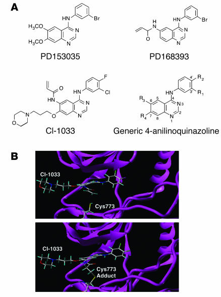
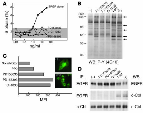
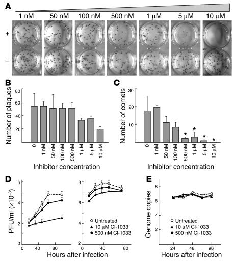
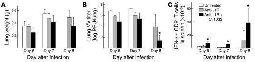
The EGF-like domain of smallpox growth factor (SPGF) targets human ErbB-1, inducing tyrosine phosphorylation of certain host cellular substrates via activation of the receptor's kinase domain and thereby facilitating viral replication. Given these findings, low molecular weight organic inhibitors of ErbB-1 kinases might function as antiviral agents against smallpox. Here we show that CI-1033 and related 4-anilinoquinazolines inhibit SPGF-induced human cellular DNA synthesis, protein tyrosine kinase activation, and c-Cbl association with ErbB-1 and resultant internalization. Infection of monkey kidney BSC-40 and VERO-E6 cells in vitro by variola strain Solaimen is blocked by CI-1033, primarily at the level of secondary viral spreading. In an in vivo lethal vaccinia virus pneumonia model, CI-1033 alone promotes survival of animals, augments systemic T cell immunity and, in conjunction with a single dose of anti-L1R intracellular mature virus particle-specific mAb, fosters virtually complete viral clearance of the lungs of infected mice by the eighth day after infection. Collectively, these findings show that chemical inhibitors of host-signaling pathways exploited by viral pathogens may represent potent antiviral therapies.


Figures

Figure 1
Figure 1
Structure of ErbB tyrosine kinase inhibitors. (A) Chemical structures of ErbB tyrosine kinase inhibitors. (B) Molecular model of CI-1033 interaction with the ErbB-1 kinase domain. The upper panel shows the model of the noncovalently bound CI-1033 in the ATP-binding site of the tyrosine kinase ErbB-1 (protein data bank: 1M17) crystal structure. The Tarceva compound was removed, and CI-1033 was manually modeled into the site. The backbone of the kinase is shown as a ribbon diagram along with the atoms of Cys773. The lower panel shows the model of the covalently bound CI-1033 in the ATP-binding site. The atoms are shown as stick models with carbon atoms colored white, oxygen atoms red, nitrogen atoms dark blue, sulfur atoms yellow, and hydrogen atoms light blue.
Figure 2
Figure 2
Functional effects of SPGF are blocked by ErbB inhibitors. (A) Inhibition of human foreskin fibroblast proliferation by ErbB inhibitors. Human fibroblasts were treated and processed as described in Methods. Percentage of cells entering S phase is plotted. Gray area represents the baseline S phase after serum starvation. (B) Inhibitors block cellular protein tyrosine phosphorylation triggered by SPGF. HeLa cells were pretreated with 50 nM inhibitors (PP2, 10 μM) at 37°C for 30 minutes and then stimulated with 50 ng/ml SPGF at 37°C for 15 minutes. Minus (–) indicates no inhibitors or SPGF, and plus (+) indicates SPGF addition only. Total cell lysates were analyzed by Western blotting (WB) with 4G10 anti-phosphotyrosine (P-Y) mAb. Arrows indicate positions of phosphorylated substrates affected by ErbB-1 inhibition. Numbers on left side of the panel indicate the molecular marker in kDa. (C) ErbB-1 internalization prevented by inhibitors. A431 cells were pretreated and processed as described in Methods. MFI is recorded by FACS. Results are representative of 3 independent experiments. The inset shows the ErbB-1 cellular distribution pattern in HeLa cells in the absence (–) or presence of SPGF. (D) Inhibitors block association of ErbB-1 and c-Cbl and subsequent ErbB-1 degradation. HeLa cells were pretreated with inhibitors and stimulated (37°C, 5 minutes) with SPGF as above. Total cell lysates were immunoprecipitated (IP) with anti-EGFR or anti–c-Cbl and subjected to Western blot with either antibody.
Figure 3
Figure 3
Prevention of secondary variola virus spreading in vitro by ErbB kinase inhibition. (A) Immunohistochemical staining of variola strain Solaimen plaques on monolayers of BSC-40 cells after 4 days in the presence (+) or absence (–) of indicated M concentrations of CI-1033. (B) Titration effect of CI-1033 on variola plaque formation. (C) Titration effect of CI-1033 on variola comet formation. Asterisks indicate statistically significant differences (P < 0.05) from 7 randomly selected control wells. (D) Time course of variola virus production (single-step growth curve) on VERO-E6 cells in the absence (untreated) or presence of indicated concentration of CI-1033 with left and right panels showing EEV and CAV titers, respectively. (E) Viral DNA genome copies from variola single-step growth curve in VERO-E6 cell in the absence (untreated) or presence of 500 nM or 10 μM CI-1033.
Figure 4
Figure 4
Rescue of mice from lethal vaccinia pneumonia by an ErbB inhibitor. (A) Mouse survival curves after i.n. vaccinia inoculation following pretreatment with CI-1033 (1 mg/mouse/day) and/or anti-L1R (200 μg i.p. 6 hours before inoculation). Note that all control mice died by day 7. Each treatment group includes a cohort of 5 animals. (B) Clinical score of mice during treatment. The score (0–4) is explained in Methods and is the mean value of the 5 mice per group. (C) Average weight of the entire lung 6 days after lethal vaccinia infection with or without indicated treatment (n = 3 per group) versus normal, uninfected lung. (D) Virus titers in lungs at day 8 after infection. Results are representative of 5 experiments.
Figure 5
Figure 5
Postexposure therapy of VV infection in the C57BL/6 pneumonia model. Mice were inoculated i.n. with 2 × 10⁴ PFU of VV, and 2 days later were treated with a single dose of mAb L1R or control mAb and/or with CI-1033, the latter given daily until termination of the experiment. Mice were sacrificed on different days after infection and examined for lung weight (A), VV PFU/lung (B), and the number of anti-CD3–induced IFN-γ–producing CD8+ T cells per spleen (C). For days 6, 7, and 8, group sizes of 3–4, 5, or 6 animals, respectively, were employed. At day 8, 2 of 6 anti–L1R-treated mice had undetectable virus, whereas 5 of 6 anti-L1R plus CI-1033–treated mice had undetectable virus. For calculation of mean titers, the lowest detectable titer possible (1 log) was used for mice without detectable virus, such that the means are actually lower values. *P < 0.05.

Comment in

References

    1. Balfour HH. Antiviral drugs. N. Engl. J. Med. 1999;340:1255–1268. - PubMed
    1. Dolin, R. 2001. Antiviral chemotherapy, excluding antiretroviral drugs. In Principles of Internal Medicine. 15th edition. E. Braunwald et al., editors. McGraw-Hill. New York, New York, USA. 1092–1100.
    1. Fauci, A.S., and Lane, H.C. 2001. Human immunodeficiency virus (HIV) disease: AIDS and related disorders. In Principles of Internal Medicine. 15th edition. E. Braunwald et al., editors. McGraw-Hill. New York, New York, USA. 1852–1913.
    1. Fenner, F., Wittek, R., and Dumbell, K.R. 1988. The orthopoxviruses. Academic Press. San Diego, California, USA. 432 pp.
    1. Henderson DA, et al. Smallpox as a biological weapon: Medical and public health management. JAMA. 1999;281:2127–2137. - PubMed

Publication types

MeSH terms