

Development of an in vivo method to investigate biomechanical and neurophysiological properties of spine facet joint capsules
- PMID: 15690211
- PMCID: PMC3489229
- DOI: 10.1007/s00586-004-0835-9
Development of an in vivo method to investigate biomechanical and neurophysiological properties of spine facet joint capsules
Abstract
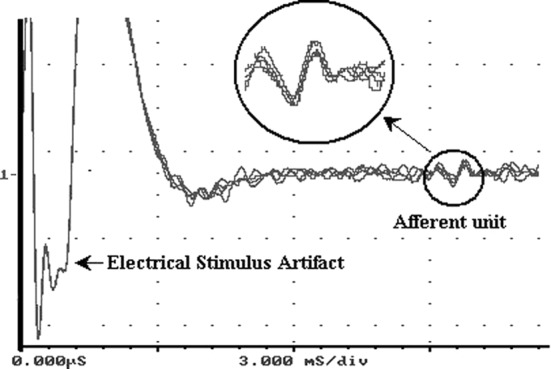
Facet joint capsules (FJC) may experience large mechanical deformation under spine motion. There has been no previous quantitative study of the relationship between capsular strain and sensory nerve activation in spine FJC in vivo. Space limitation in the cervical spine makes such a study difficult, as the facet joint must be loaded while simultaneously monitoring nerve discharge from nerve roots immediately adjacent to the loaded tissue. A new methodology was developed to investigate biomechanical and neurophysiological properties of spine facet joint capsules in vivo. The method incorporated a custom-fabricated testing frame for facet joint loading, a stereoimaging system, and a template-matching technique to obtain single afferent response. It was tested by loading goat C5-C6 FJC in vivo with simultaneous nerve root recordings and 3D strain tracking of the capsules. Preliminary data showed that 18 of 23 afferents (78.3%) were found to be mechanosensitive to tensile stretch, and five were not responsive, even under tensile load as high as 27.5 N. Mechanosensitive afferents in goat capsules had tensile strain thresholds of 0.119+/-0.080. Neural responses of all mechanosensitive units showed statistically significant correlations (all P<<0.05) with both capsular load (r(2)=0.744+/-0.109) and local strain (r(2)=0.868+/-0.088). This method enables the investigation of the correlation between tissue load, deformation and neural responses of mechanoreceptors in spine facet joint capsules, and can be adapted to investigate tissue loading and neural response of other soft tissues.
Figures


Similar articles
-
Neurophysiological and biomechanical characterization of goat cervical facet joint capsules.J Orthop Res. 2005 Jul;23(4):779-87. doi: 10.1016/j.orthres.2005.01.002. Epub 2005 Feb 19. J Orthop Res. 2005. PMID: 16022990
-
Recording of neural activity from goat cervical facet joint capsule using custom-designed miniature electrodes.Spine (Phila Pa 1976). 2005 Jun 15;30(12):1367-72. doi: 10.1097/01.brs.0000166193.39389.21. Spine (Phila Pa 1976). 2005. PMID: 15959364
-
Strain and load thresholds for cervical muscle recruitment in response to quasi-static tensile stretch of the caprine C5-C6 facet joint capsule.J Electromyogr Kinesiol. 2009 Dec;19(6):e387-94. doi: 10.1016/j.jelekin.2009.01.002. Epub 2009 Feb 14. J Electromyogr Kinesiol. 2009. PMID: 19223204
-
Pain generation in lumbar and cervical facet joints.J Bone Joint Surg Am. 2006 Apr;88 Suppl 2:63-7. doi: 10.2106/JBJS.E.01411. J Bone Joint Surg Am. 2006. PMID: 16595446 Review.
-
Mechanoreceptor endings in human thoracic and lumbar facet joints.Spine (Phila Pa 1976). 1998 Jan 15;23(2):168-73. doi: 10.1097/00007632-199801150-00004. Spine (Phila Pa 1976). 1998. PMID: 9474721 Review.
Cited by
-
The role of tissue damage in whiplash-associated disorders: discussion paper 1.Spine (Phila Pa 1976). 2011 Dec 1;36(25 Suppl):S309-15. doi: 10.1097/BRS.0b013e318238842a. Spine (Phila Pa 1976). 2011. PMID: 22020601 Free PMC article. Review.
-
Applications of SPECT/CT in the Evaluation of Spinal Pathology: A Review.Int J Spine Surg. 2024 Mar 4;18(1):9-23. doi: 10.14444/8552. Int J Spine Surg. 2024. PMID: 38050030 Free PMC article.
-
Tensile stretching of cervical facet joint capsule and related axonal changes.Eur Spine J. 2008 Apr;17(4):556-63. doi: 10.1007/s00586-007-0562-0. Epub 2007 Dec 14. Eur Spine J. 2008. PMID: 18080147 Free PMC article.
-
Whiplash causes increased laxity of cervical capsular ligament.Clin Biomech (Bristol). 2008 Feb;23(2):159-65. doi: 10.1016/j.clinbiomech.2007.09.003. Epub 2007 Oct 23. Clin Biomech (Bristol). 2008. PMID: 17959284 Free PMC article.
-
Neck ligament strength is decreased following whiplash trauma.BMC Musculoskelet Disord. 2006 Dec 21;7:103. doi: 10.1186/1471-2474-7-103. BMC Musculoskelet Disord. 2006. PMID: 17184536 Free PMC article.
References
-
- Avramov AI, Cavanaugh JM, Ozaktay CA, Getchell TV, King AI. The effects of controlled mechanical loading on group-II, III, and IV afferent units from the lumbar facet joint and surrounding tissue. An in vitro study. J Bone Joint Surg Am. 1992;74:1464–1471. - PubMed
-
- Bogduk N. The clinical anatomy of the cervical dorsal rami. Spine. 1982;7:319–330. - PubMed
-
- Bogduk N, Marsland A. The cervical zygapophysial joints as a source of neck pain. Spine. 1988;13:610–617. - PubMed
-
- Brantigan JW, McAfee PC, Cunningham BW, Wang H, Orbegoso CM. Interbody lumbar fusion using a carbon fiber cage implant versus allograft bone. An investigational study in the Spanish goat. Spine. 1994;19:1436–1444. - PubMed
Publication types
MeSH terms
LinkOut - more resources
Full Text Sources
Miscellaneous
