. 2005 Jul;204(1):210-8.
doi: 10.1002/jcp.20285.

Abstrakt interacts with and regulates the expression of sorting nexin-2

Affiliations

Abstrakt interacts with and regulates the expression of sorting nexin-2

Mohammad Abdul-Ghani et al. J Cell Physiol. 2005 Jul.

Abstract

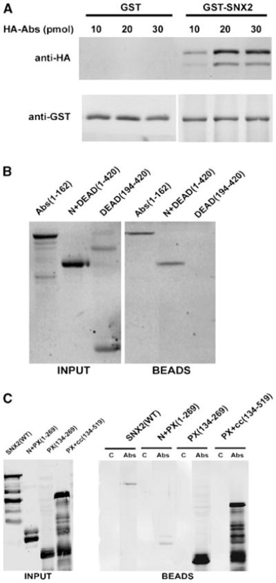
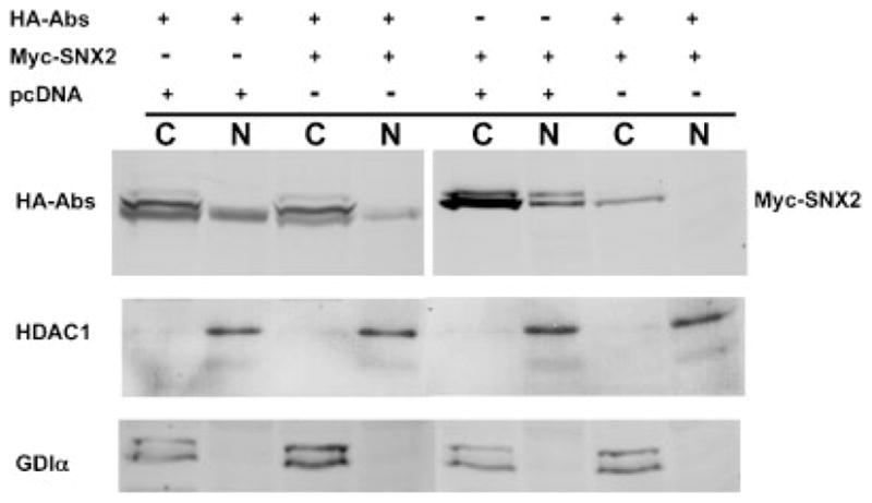
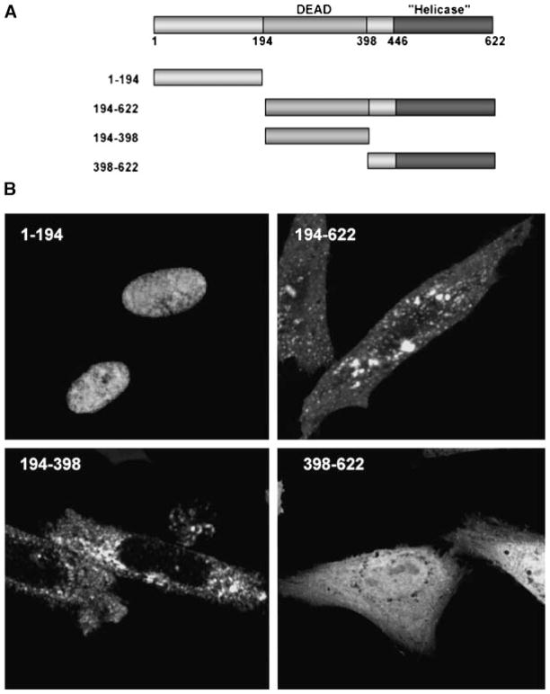
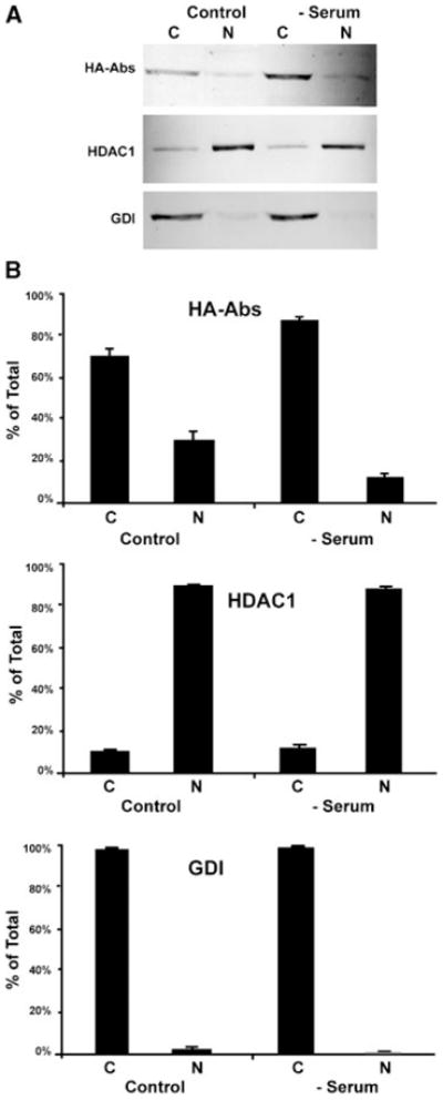
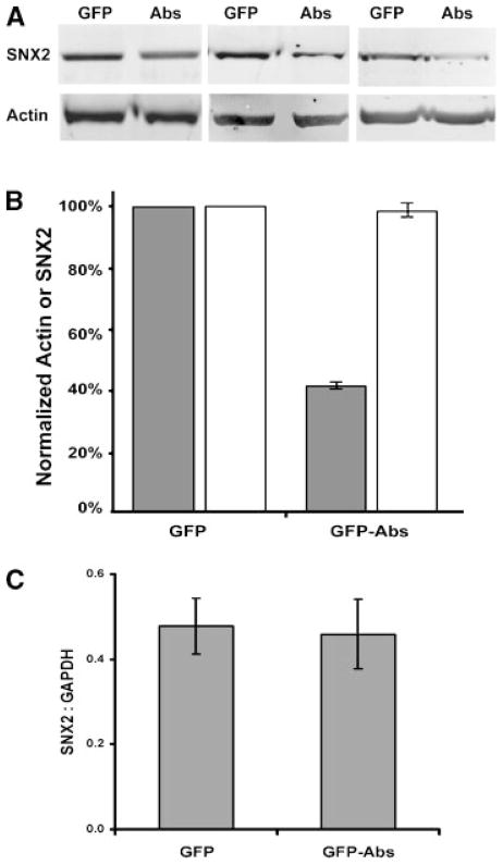
Protein sorting through vesicular compartments is highly regulated to maintain the integrity and signaling of intracellular organelles in eukaryotic cells. Sorting Nexin-2 (SNX2) is involved in protein sorting in the trans-Golgi network, endosome, and/or lysosome compartments, with loss of function leading to defect in protein sorting and stress on organelles. To investigate the function of SNX2, we have identified the DEAD-box helicase Abstrakt (Abs) as an SNX2-interacting protein. The N-terminal domain of Abs interacts with the phox homology (PX) domain of SNX2 suggesting that PX domains may also participate in protein-protein interaction. Interestingly, both proteins undergo nucleocytoplasmic shuttling, and this process is responsive to serum withdrawal for Abs. Finally, expression of Abs reduced the cellular expression of SNX2 without altering its steady state mRNA levels. This unexpected interaction provides a novel mechanism whereby expression of proteins involved in membrane trafficking could be regulated by an RNA helicase.


Figures

Fig. 1
Fig. 1
In vitro pulldown of Abstrakt (Abs) and sorting nexin-2 (SNX2). A: Increasing amounts of recombinant hemagglutinin (HA)-Abs was added to 5 pmol of GST-SNX2 or GST pre-bound to glutathione-agarose beads. Proteins bound to the beads were analyzed with anti-HA (top part) and anti-GST (bottom part) antibodies. B: Various HA-Abs deletion fragments (20 pmol each) were added to GST-SNX2 bound to glutathione-Sepharose beads. Numbers in parentheses indicate the inclusive range of amino acids in each Abs fragment. HA-Abs in the input (left part) and bound to the beads (right part) were analyzed with anti-HA. Fraction of the Abs DEAD(194-420) fragment often appear as a SDS-resistant dimer. C: Various FLAG-SNX2 fragments (labeled input, left part) were added to control Sepharose 4B beads (lanes labeled C, right part) or to equal volume of Abs immobilized on Sepharose 4B (lanes labeled Abs, right part). FLAG-SNX2 associated with the beads was detected with anti-FLAG antibodies.
Fig. 2
Fig. 2
Cellular localization of GFP-Abs and Myc-SNX2. A, B: Representative images of Chinese hamster ovary (CHO) transfected with GFP-Abs. Most cells (~90%) exhibited a prominent nuclear signal (A) while some (~10%) exhibited both nuclear and cytosolic localization. C: CHO transfected with Myc-SNX2 showed a uniformly punctate distribution consistent with the endocytic compartment. DF CHO co-transfected with GFP-Abs and Myc-SNX2 showing the cytosolic distribution of GFP-Abs (D), Myc-SNX2 (E), and merged (F) images.
Fig. 3
Fig. 3
Cell fusion assay of GFP-Abs and nuclear localization of SNX2. CHO were transfected with GFP-Abs and fused with un-transfected CHO cells. All multi-nucleated cells exhibited a fluorescent nuclear signal (A, B). CHO transfected with Myc-SNX2 showed normal cytoplasmic punctate staining (C) but with significant nuclear localization when treated with the nuclear export blocker Leptomycin B (LMB) (D).
Fig. 4
Fig. 4
Fluorescence recovery after photobleaching (FRAP) analysis of GFP-Abs in fused, multi-nucleated CHO. CHO cells expressing GFP-Abs were fused with un-transfected cells. A: One of the nuclei was photobleached (arrowhead) and fluorescence recovery was monitored at 10 min intervals for 90 min as indicated on the lower right hand corner of each part. B: FRAP of a nucleus (arrowhead) in a multi-nucleated cell pre-treated with the nuclear export inhibitor LMB showing no fluorescence recovery (only the 90 min recovery time point is shown).
Fig. 5
Fig. 5
Nuclear and cytoplasmic fractionation of HA-Abs and Myc-SNX2. CHO were transfected with a combination of HA-Abs, Myc-SNX2, and empty pcDNA3.1(+) vector control and fractionated using the NE-PER kit. One quarter of each fractions were analyzed by Western immunoblot with anti-HA and anti-Myc antibodies. Histone deacetylase-1 (HDAC-1) and GDP-dissociation inhibitor (GDI)α were used as nuclear and cytosolic controls, respectively. Lanes indicated with C represents the cytosolic fraction and N the nuclear fraction.
Fig. 6
Fig. 6
Identification of the NLS within Abs. A: Schematic diagram of full length and truncated Abs with the DEAD-box, helicase domain, and residue number as indicated. The various Abs fragments were inserted into the C-terminus of GFP. B: Representative images of transfected CHO cells. The N-terminal 194 amino acids targeted GFP to the nucleus. Deletion of this domain in Abs(194-622) and Abs(194-398) resulted in punctate cytoplasmic staining and exclusion from the nucleus. Fusion with the helicase domain in Abs(398-622) resulted in uniform nuclear and cytoplasmic staining.
Fig. 7
Fig. 7
Serum withdrawal alters cellular localization of Abs and SNX2. HeLa cells were transfected with HA-Abs. After 24 h, fetal bovine serum (FBS) was withdrawn for 4 h (serum) prior to NE-PER nuclear fractionation while control cells were maintained in 5% FBS for the duration. A: Shows a representative Western immunoblot of the NE-PER fractions probed with anti-HA, HDAC, and GDI antibodies, as indicated. Each lane represents one quarter of the cytoplasmic (C) and nuclear (N) fractions. B: Quantitative analysis of the relative distribution of Abs, HDAC-1, and GDI in the NE-PER fractions indicate a shift in Abs from nuclear to cytoplasmic pool upon serum withdrawal (mean ± SEM; n = 4). The nuclear and cytoplasmic fractions were indicated by the nuclear and cytoplasmic localized HDAC1 and GDI, respectively.
Fig. 8
Fig. 8
Expression of GFP-Abs down regulates endogenous SNX2. HeLa transiently transfected with GFP control or GFP-Abs were enriched by fluorescence activated cell sorter (FACS). A: Crude lysate (15 μg) from three experiments were analyzed by Western immunoblot with anti-SNX2 antibodies. The blots were also probed for actin. B: Signal intensities were quantified using ImageQuant 5.2 Software (Amersham Biosciences, Piscataway, NJ). SNX2 (shaded bar) and actin (clear bar) values from GFP-Abs expressing cells were expressed as a percentage of the GFP controls. C: Quantitative RT-PCR analysis of HeLa cells stably transfected with GFP or GFP-Abs. SNX2 mRNA values normalized to that of GAPDH (mean ± SEM; n = 3) were from three independent stable lines.

References

    1. Abram CL, Seals DF, Pass I, Salinsky D, Maurer L, Roth TM, Courtneidge SA. The adaptor protein fish associates with members of the ADAMs family and localizes to podosomes of Src-transformed cells. J Biol Chem. 2003;278(19):16844–16851. - PubMed
    1. Coda L, Salcini AE, Confalonieri S, Pelicci G, Sorkina T, Sorkin A, Pelicci PG, Di Fiore PP. Eps15R is a tyrosine kinase substrate with characteristics of a docking protein possibly involved in coated pits-mediated internalization. J Biol Chem. 1998;273(5):3003–3012. - PubMed
    1. Conibear E, Stevens TH. Multiple sorting pathways between the late Golgi and the vacuole in yeast. Biochim Biophys Acta. 1998;1404(1–2):211–230. - PubMed
    1. Cozier GE, Carlton J, McGregor AH, Gleeson PA, Teasdale RD, Mellor H, Cullen PJ. The phox homolosgy (PX) domain-dependent, 3-phosphoinositide-mediated association of sorting Nexin-1 with an early sorting endosomal compartment is required for its ability to regulate epidermal growth factor receptor degradation. J Biol Chem. 2002;277(50):48730–48736. - PubMed
    1. Deloche O, de la Cruz J, Kressler D, Doere M, Linder P. A membrane transport defect leads to a rapid attenuation of translation initiation in Saccharomyces cerevisiae. Mol Cell. 2004;13(3):357–366. - PubMed

Publication types

MeSH terms

Grants and funding

LinkOut - more resources