

Automatic lesion boundary detection in dermoscopy images using gradient vector flow snakes
- PMID: 15691255
- PMCID: PMC3184888
- DOI: 10.1111/j.1600-0846.2005.00092.x
Automatic lesion boundary detection in dermoscopy images using gradient vector flow snakes
Abstract
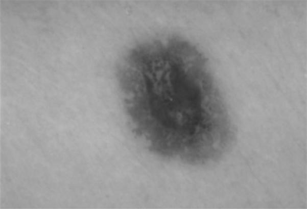
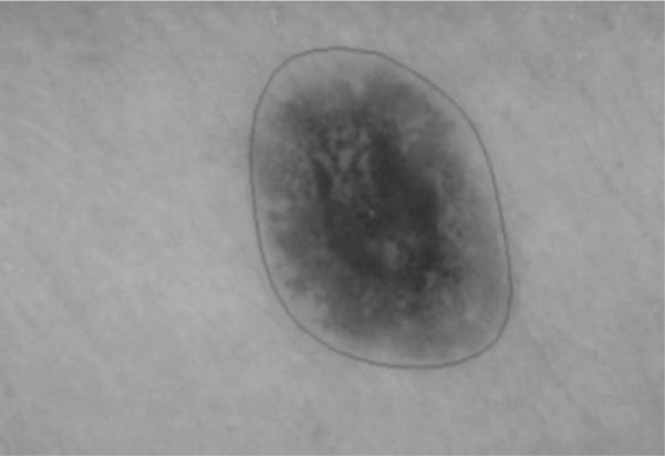
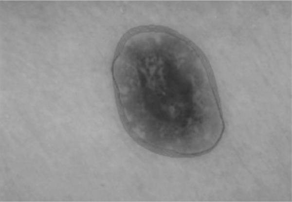
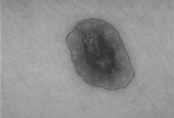
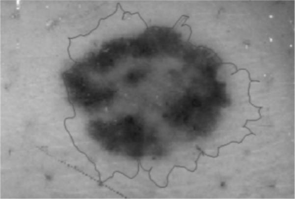
Background: Malignant melanoma has a good prognosis if treated early. Dermoscopy images of pigmented lesions are most commonly taken at x 10 magnification under lighting at a low angle of incidence while the skin is immersed in oil under a glass plate. Accurate skin lesion segmentation from the background skin is important because some of the features anticipated to be used for diagnosis deal with shape of the lesion and others deal with the color of the lesion compared with the color of the surrounding skin.
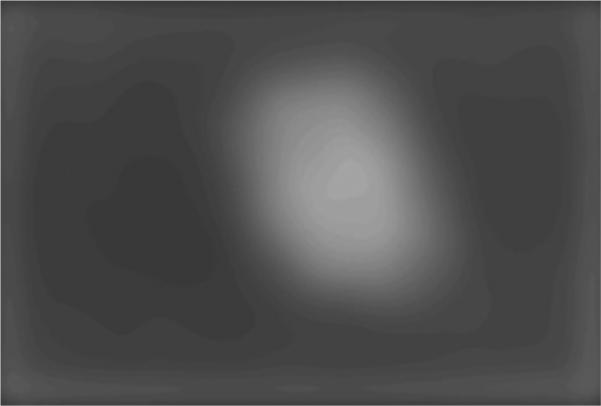
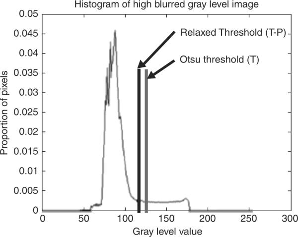
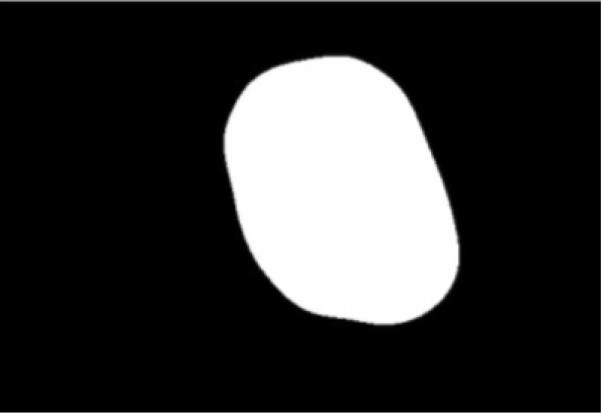
Methods: In this research, gradient vector flow (GVF) snakes are investigated to find the border of skin lesions in dermoscopy images. An automatic initialization method is introduced to make the skin lesion border determination process fully automated.
Results: Skin lesion segmentation results are presented for 70 benign and 30 melanoma skin lesion images for the GVF-based method and a color histogram analysis technique. The average errors obtained by the GVF-based method are lower for both the benign and melanoma image sets than for the color histogram analysis technique based on comparison with manually segmented lesions determined by a dermatologist.
Conclusions: The experimental results for the GVF-based method demonstrate promise as an automated technique for skin lesion segmentation in dermoscopy images.
Figures

Similar articles
-
Gradient vector flow with mean shift for skin lesion segmentation.Comput Med Imaging Graph. 2011 Mar;35(2):121-7. doi: 10.1016/j.compmedimag.2010.08.002. Epub 2010 Sep 15. Comput Med Imaging Graph. 2011. PMID: 20832242
-
Modified watershed technique and post-processing for segmentation of skin lesions in dermoscopy images.Comput Med Imaging Graph. 2011 Mar;35(2):116-20. doi: 10.1016/j.compmedimag.2010.09.006. Epub 2010 Oct 20. Comput Med Imaging Graph. 2011. PMID: 20970307 Free PMC article.
-
Approximate lesion localization in dermoscopy images.Skin Res Technol. 2009 Aug;15(3):314-22. doi: 10.1111/j.1600-0846.2009.00357.x. Skin Res Technol. 2009. PMID: 19624428 Free PMC article.
-
Lesion border detection in dermoscopy images.Comput Med Imaging Graph. 2009 Mar;33(2):148-53. doi: 10.1016/j.compmedimag.2008.11.002. Epub 2009 Jan 3. Comput Med Imaging Graph. 2009. PMID: 19121917 Free PMC article. Review.
-
Dermoscopy, with and without visual inspection, for diagnosing melanoma in adults.Cochrane Database Syst Rev. 2018 Dec 4;12(12):CD011902. doi: 10.1002/14651858.CD011902.pub2. Cochrane Database Syst Rev. 2018. PMID: 30521682 Free PMC article.
Cited by
-
Skin lesion classification using multi-resolution empirical mode decomposition and local binary pattern.PLoS One. 2022 Sep 20;17(9):e0274896. doi: 10.1371/journal.pone.0274896. eCollection 2022. PLoS One. 2022. PMID: 36126072 Free PMC article.
-
Artificial Intelligence-Based Approaches to Reflectance Confocal Microscopy Image Analysis in Dermatology.J Clin Med. 2022 Jan 14;11(2):429. doi: 10.3390/jcm11020429. J Clin Med. 2022. PMID: 35054123 Free PMC article. Review.
-
Developing a Recognition System for Diagnosing Melanoma Skin Lesions Using Artificial Intelligence Algorithms.Comput Math Methods Med. 2021 May 15;2021:9998379. doi: 10.1155/2021/9998379. eCollection 2021. Comput Math Methods Med. 2021. PMID: 34055044 Free PMC article.
-
Fuzzy logic color detection: Blue areas in melanoma dermoscopy images.Comput Med Imaging Graph. 2014 Jul;38(5):403-10. doi: 10.1016/j.compmedimag.2014.03.007. Epub 2014 Apr 3. Comput Med Imaging Graph. 2014. PMID: 24786720 Free PMC article.
-
Improving Skin Lesion Segmentation with Self-Training.Cancers (Basel). 2024 Mar 11;16(6):1120. doi: 10.3390/cancers16061120. Cancers (Basel). 2024. PMID: 38539454 Free PMC article.
References
-
- Saphier J, Dermatoskopie D, Mitteilung I. Arch Dermatitis Syphillis. 1921;128:1–19.
-
- Steiner A, Pehamberger H, Wolff K. Improvement of diagnostic accuracy in pigmented skin lesions by epiluminescent light microscopy. J Am Acad Dermatol. 1987;17:571–573. - PubMed
-
- Nachbar F, Stolz W, Merkle T, et al. The ABCD rule of dermatoscopy. J Am Acad Dermatol. 1994;30:551–559. - PubMed
-
- Binder M, Schwarz M, Winkler A, Steiner A, Kaider A, Wolff K, Pehamberger H. Epiluminescence microscopy. A useful tool for the diagnosis of pigmented skin lesions for formally trained dermatologists. Arch Dermatol. 1995;131:286–291. - PubMed
-
- Cristofolini M, Zumiani G, Bauer P, Cristofolini P, Boi S, Micciolo R. Dermatoscopy usefulness in the differential diagnosis of cutaneous pigmentary lesions. Melanoma Res. 1994;4:391–394. - PubMed
Publication types
MeSH terms
Grants and funding
LinkOut - more resources
Full Text Sources
Other Literature Sources
Medical
