

Low-temperature growth of Shewanella oneidensis MR-1
- PMID: 15691935
- PMCID: PMC546687
- DOI: 10.1128/AEM.71.2.811-816.2005
Low-temperature growth of Shewanella oneidensis MR-1
Abstract
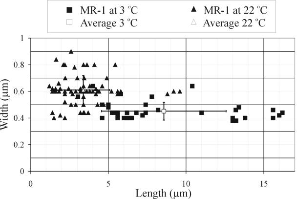
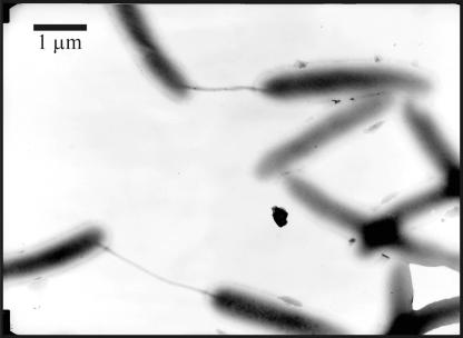
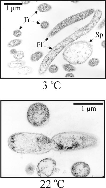
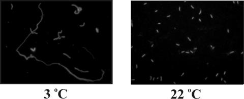
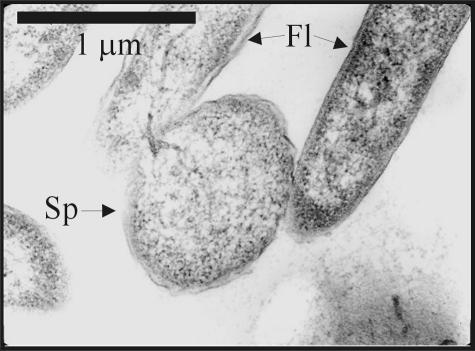
Shewanella oneidensis MR-1 is a mesophilic bacterium with a maximum growth temperature of approximately 35 degrees C but the ability to grow over a wide range of temperatures, including temperatures near zero. At room temperature ( approximately 22 degrees C) MR-1 grows with a doubling time of about 40 min, but when moved from 22 degrees C to 3 degrees C, MR-1 cells display a very long lag phase of more than 100 h followed by very slow growth, with a doubling time of approximately 67 h. In comparison to cells grown at 22 degrees C, the cold-grown cells formed long, motile filaments, showed many spheroplast-like structures, produced an array of proteins not seen at higher temperature, and synthesized a different pattern of cellular lipids. Frequent pilus-like structures were observed during the transition from 3 to 22 degrees C.
Figures
Similar articles
-
Characterization of the lipopolysaccharides and capsules of Shewanella spp.Appl Environ Microbiol. 2002 Sep;68(9):4653-7. doi: 10.1128/AEM.68.9.4653-4657.2002. Appl Environ Microbiol. 2002. PMID: 12200327 Free PMC article.
-
Evaluation of the effects of various culture conditions on Cr(VI) reduction by Shewanella oneidensis MR-1 in a novel high-throughput mini-bioreactor.Biotechnol Bioeng. 2006 Sep 5;95(1):176-84. doi: 10.1002/bit.21002. Biotechnol Bioeng. 2006. PMID: 16732598
-
Improving solubility of Shewanella oneidensis MR-1 and Clostridium thermocellum JW-20 proteins expressed into Esherichia coli.J Proteome Res. 2005 Nov-Dec;4(6):1942-51. doi: 10.1021/pr050108j. J Proteome Res. 2005. PMID: 16335938
-
Global transcriptome analysis of the cold shock response of Shewanella oneidensis MR-1 and mutational analysis of its classical cold shock proteins.J Bacteriol. 2006 Jun;188(12):4560-9. doi: 10.1128/JB.01908-05. J Bacteriol. 2006. PMID: 16740962 Free PMC article.
-
Methods for imaging Shewanella oneidensis MR-1 nanofilaments.J Microbiol Methods. 2010 Aug;82(2):187-91. doi: 10.1016/j.mimet.2010.05.011. Epub 2010 Jun 1. J Microbiol Methods. 2010. PMID: 20561956
Cited by
-
The Contribution of High-Order Metabolic Interactions to the Global Activity of a Four-Species Microbial Community.PLoS Comput Biol. 2016 Sep 13;12(9):e1005079. doi: 10.1371/journal.pcbi.1005079. eCollection 2016 Sep. PLoS Comput Biol. 2016. PMID: 27623159 Free PMC article.
-
The psychrophilic lifestyle as revealed by the genome sequence of Colwellia psychrerythraea 34H through genomic and proteomic analyses.Proc Natl Acad Sci U S A. 2005 Aug 2;102(31):10913-8. doi: 10.1073/pnas.0504766102. Epub 2005 Jul 25. Proc Natl Acad Sci U S A. 2005. PMID: 16043709 Free PMC article.
-
Host genetic selection for cold tolerance shapes microbiome composition and modulates its response to temperature.Elife. 2018 Nov 20;7:e36398. doi: 10.7554/eLife.36398. Elife. 2018. PMID: 30454554 Free PMC article.
-
Whole-Cell Biosensor for Iron Monitoring as a Potential Tool for Safeguarding Biodiversity in Polar Marine Environments.Mar Drugs. 2024 Jun 28;22(7):299. doi: 10.3390/md22070299. Mar Drugs. 2024. PMID: 39057408 Free PMC article. Review.
-
In vivo characterization of electroactive biofilms inside porous electrodes with MR Imaging.RSC Adv. 2022 Jun 15;12(28):17784-17793. doi: 10.1039/d2ra01162j. eCollection 2022 Jun 14. RSC Adv. 2022. PMID: 35765339 Free PMC article.
References
-
- Anderson, N. G., and N. L. Anderson. 1978. Analytical techniques for cell fractions. 21. 2-dimensional analysis of serum and tissue proteins-multiple isoelectric-focusing. Anal. Biochem. 85:331-340. - PubMed
-
- Anderson, N. L., and N. G. Anderson. 1978. Analytical techniques for cell fractions. 22. 2-dimensional analysis of serum and tissue proteins-multiple gradient-slab gel-electrophoresis. Anal. Biochem. 85:341-354. - PubMed
Publication types
MeSH terms
Substances
LinkOut - more resources
Full Text Sources
Other Literature Sources
Molecular Biology Databases
