Metabolism of bismuth subsalicylate and intracellular accumulation of bismuth by Fusarium sp. strain BI
- PMID: 15691943
- PMCID: PMC546758
- DOI: 10.1128/AEM.71.2.876-882.2005
Metabolism of bismuth subsalicylate and intracellular accumulation of bismuth by Fusarium sp. strain BI
Abstract
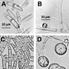
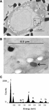
Enrichment cultures were conducted using bismuth subsalicylate as the sole source of carbon and activated sludge as the inoculum. A pure culture was obtained and identified as a Fusarium sp. based on spore morphology and partial sequences of 18S rRNA, translation elongation factor 1-alpha, and beta-tubulin genes. The isolate, named Fusarium sp. strain BI, grew to equivalent densities when using salicylate or bismuth subsalicylate as carbon sources. Bismuth nitrate at concentrations of up to 200 muM did not limit growth of this organism on glucose. The concentration of soluble bismuth in suspensions of bismuth subsalicylate decreased during growth of Fusarium sp. strain BI. Transmission electron microscopy and energy-dispersive spectroscopy revealed that the accumulated bismuth was localized in phosphorus-rich granules distributed in the cytoplasm and vacuoles. Long-chain polyphosphates were extracted from fresh biomass grown on bismuth subsalicylate, and inductively coupled plasma optical emission spectrometry showed that these fractions also contained high concentrations of bismuth. Enzyme activity assays of crude extracts of Fusarium sp. strain BI showed that salicylate hydroxylase and catechol 1,2-dioxygenase were induced during growth on salicylate, indicating that this organism degrades salicylate by conversion of salicylate to catechol, followed by ortho cleavage of the aromatic ring. Catechol 2,3-dioxygenase activity was not detected. Fusarium sp. strain BI grew with several other aromatic acids as carbon sources: benzoate, 3-hydroxybenzoate, 4-hydroxybenzoate, gentisate, d-mandelate, l-phenylalanine, l-tyrosine, phenylacetate, 3-hydroxyphenylacetate, 4-hydroxyphenylacetate, and phenylpropionate.
Figures


Similar articles
-
Salicylate absorption from a bismuth subsalicylate preparation.Clin Pharmacol Ther. 1981 Jun;29(6):788-92. doi: 10.1038/clpt.1981.112. Clin Pharmacol Ther. 1981. PMID: 7226711
-
Absorption of salicylate and bismuth from a bismuth subsalicylate--containing compound (Pepto-Bismol).J Pediatr. 1981 Oct;99(4):654-6. doi: 10.1016/s0022-3476(81)80285-5. J Pediatr. 1981. PMID: 7277115 No abstract available.
-
Bismuth subsalicylate reduces peptic injury of the oesophagus in rabbits.Gut. 1990 Jan;31(1):11-6. doi: 10.1136/gut.31.1.11. Gut. 1990. PMID: 2108076 Free PMC article.
-
Bismuth subsalicylate: history, chemistry, and safety.Rev Infect Dis. 1990 Jan-Feb;12 Suppl 1:S3-8. doi: 10.1093/clinids/12.supplement_1.s3. Rev Infect Dis. 1990. PMID: 2406853 Review.
-
Alkyl derivatives of bismuth in environmental and biological media.Met Ions Life Sci. 2010;7:303-18. doi: 10.1039/9781849730822-00303. Epub 2010 Jan 30. Met Ions Life Sci. 2010. PMID: 20877811 Review.
Cited by
-
A new mercury-accumulating Mucor hiemalis strain EH8 from cold sulfidic spring water biofilms.Microbiologyopen. 2016 Oct;5(5):763-781. doi: 10.1002/mbo3.368. Epub 2016 May 13. Microbiologyopen. 2016. PMID: 27177603 Free PMC article.
-
Complementary Proteomics, Genomics approaches identifies potential pathogenicity/virulence factors in Tilletia indica induced under the influence of host factor.Sci Rep. 2019 Jan 24;9(1):553. doi: 10.1038/s41598-018-37810-1. Sci Rep. 2019. PMID: 30679765 Free PMC article.
-
Warming Scenarios and Phytophthora cinnamomi Infection in Chestnut (Castanea sativa Mill.).Plants (Basel). 2023 Jan 26;12(3):556. doi: 10.3390/plants12030556. Plants (Basel). 2023. PMID: 36771639 Free PMC article.
-
Hydrogel-based dressings in the treatment of partial thickness experimentally induced burn wounds in rats.Acta Cir Bras. 2022 Jul 1;37(4):e370401. doi: 10.1590/acb370401. eCollection 2022. Acta Cir Bras. 2022. PMID: 35792743 Free PMC article.
-
Polyphosphate in Lactobacillus and Its Link to Stress Tolerance and Probiotic Properties.Front Microbiol. 2018 Sep 7;9:1944. doi: 10.3389/fmicb.2018.01944. eCollection 2018. Front Microbiol. 2018. PMID: 30245671 Free PMC article.
References
-
- Altschul, S. F., W. Gish, W. Miller, E. W. Myers, and D. J. Lipman. 1990. Basic local alignment search tool. J. Mol. Biol. 215:403-410. - PubMed
-
- Barz, W. 1971. Über den abbau aromatischer verbindungen durch Fusarium oxysporum schlecht. Arch. Mikrobiol. 78:341-352. - PubMed
-
- Bergmeyer, H. U., M. Grassl, and H.-E. Walter. 1983. Reagents for enzymatic analysis, p. 222-223. In H. U. Bergmeyer, J. Bergmeyer, and M. Grassl (ed.), Methods of enzymatic analysis, 3rd ed., vol. 2. Verlag Chemie, Weinheim, Germany.
Publication types
MeSH terms
Substances
Associated data
- Actions
- Actions
- Actions
Grants and funding
LinkOut - more resources
Full Text Sources
Molecular Biology Databases

