Review
. 2005;6(2):209.
doi: 10.1186/gb-2005-6-2-209. Epub 2005 Feb 1.

The vascular endothelial growth factor (VEGF) family: angiogenic factors in health and disease

Affiliations
Review

The vascular endothelial growth factor (VEGF) family: angiogenic factors in health and disease

David I R Holmes et al. Genome Biol. 2005.

Abstract

Vascular endothelial growth factors (VEGFs) are a family of secreted polypeptides with a highly conserved receptor-binding cystine-knot structure similar to that of the platelet-derived growth factors. VEGF-A, the founding member of the family, is highly conserved between animals as evolutionarily distant as fish and mammals. In vertebrates, VEGFs act through a family of cognate receptor kinases in endothelial cells to stimulate blood-vessel formation. VEGF-A has important roles in mammalian vascular development and in diseases involving abnormal growth of blood vessels; other VEGFs are also involved in the development of lymphatic vessels and disease-related angiogenesis. Invertebrate homologs of VEGFs and VEGF receptors have been identified in fly, nematode and jellyfish, where they function in developmental cell migration and neurogenesis. The existence of VEGF-like molecules and their receptors in simple invertebrates without a vascular system indicates that this family of growth factors emerged at a very early stage in the evolution of multicellular organisms to mediate primordial developmental functions.


Figures
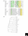
Figure 1
Figure 1
Comparison of human VEGFs with PDGFs and related sequences from Drosophila and Orf virus. Abbreviations: h, human; dm, Drosophila melanogaster, ov, Orf virus. (a) An alignment of the deduced amino-acid sequences of the VEGF/PDGF homology domain (VHD) from various human, Drosophila and Orf virus VEGFs and PGDFs. {online only} Sequence data were obtained from the GenBank and SwissProt databases; the multiple alignment was generated using MultAlin and further optimized manually. Residues that are conserved in at least 50% of the aligned sequences are shaded in green; those fully conserved are in yellow. The eight cysteine residues that constitute the cystine-knot structure [12] are denoted by asterisks below the sequences. (b) Predicted evolutionary relationships between human, Drosophila and Orf virus VEGFs and PDGFs. VHD sequences from (a) were aligned using ClustalW and the neighbor-joining method was used to construct a phylogenetic tree with TreeView. Branch lengths are proportional to the estimated evolutionary distance between protein sequences.
Figure 2
Figure 2
Gene organization and encoded functional domains of the human VEGF genes and related genes from Drosophila. Exons, represented by boxes, are numbered and the length of coding sequence in each is marked below in base-pairs. Start (ATG) and stop (TAA, TAG, TGA) codons are marked, and the length of each encoded unprocessed polypeptide including the signal peptide (in amino-acid residues) is indicated in parentheses. Exons are drawn to scale, except for the last exon of hVEGF-A, which is longer than 1 kilobase (kb). Introns, represented by horizontal lines, are not drawn to scale. Alternative exons and splicing patterns are not shown, with the exception of hVEGF-B, in which isoforms result from alternative splicing of exon 6 [23]. Arrows represent proteolytic cleavage sites. Abbreviations: 3', 3' untranslated region (UTR); 5', 5' UTR; CP, region encoding the carboxy-terminal propeptide domain; H, encodes the heparin-binding domain; N, encodes the NRP1/heparin-binding domain; NP, encodes the amino-terminal propeptide domain; SP, signal peptide; VHD, encodes the VEGF/PDGF homology domain. Information was compiled from published literature [14-16,22,23,59-61] and the Entrez Gene, RefSeq, GenBank and SwissProt databases.

References

    1. Ferrara N, Gerber HP, LeCouter J. The biology of VEGF and its receptors. Nat Med. 2003;9:669–676. doi: 10.1038/nm0603-669. A concise review of the role of VEGF-A and its receptors in biology and disease. - DOI - PubMed
    1. Senger DR, Galli SJ, Dvorak AM, Perruzzi CA, Harvey VS, Dvorak HF. Tumor cells secrete a vascular permeability factor that promotes accumulation of ascites fluid. Science. 1983;219:983–985. The initial discovery of a secreted VPF with the characteristics of VEGF-A. - PubMed
    1. Leung DW, Cachianes G, Kuang WJ, Goeddel DV, Ferrara N. Vascular endothelial growth factor is a secreted angiogenic mitogen. Science. 1989;246:1306–1309. This and [4] are the first reports of the cDNA cloning of VEGF-A. - PubMed
    1. Keck PJ, Hauser SD, Krivi G, Sanzo K, Warren T, Feder J, Connolly DT. Vascular permeability factor, an endothelial cell mitogen related to PDGF. Science. 1989;246:1309–1312. See [3]. - PubMed
    1. Li X, Eriksson U. Novel VEGF family members: VEGF-B, VEGF-C and VEGF-D. Int J Biochem Cell Biol. 2001;33:421–426. doi: 10.1016/S1357-2725(01)00027-9. A review of the mammalian VEGF family. - DOI - PubMed

Publication types

MeSH terms

Substances

LinkOut - more resources