Review
. 2005 Jun 15;171(12):1328-42.
doi: 10.1164/rccm.200408-1036SO. Epub 2005 Feb 1.

Cellular stress failure in ventilator-injured lungs

Affiliations
Review

Cellular stress failure in ventilator-injured lungs

Nicholas E Vlahakis et al. Am J Respir Crit Care Med. .

Abstract

The clinical and experimental literature has unequivocally established that mechanical ventilation with large tidal volumes is injurious to the lung. However, uncertainty about the micromechanics of injured lungs and the numerous degrees of freedom in ventilator settings leave many unanswered questions about the biophysical determinants of lung injury. In this review we focus on experimental evidence for lung cells as injury targets and the relevance of these studies for human ventilator-associated lung injury. In vitro, the stress-induced mechanical interactions between matrix and adherent cells are important for cellular remodeling as a means for preventing compromise of cell structure and ultimately cell injury or death. In vivo, these same principles apply. Large tidal volume mechanical ventilation results in physical breaks in alveolar epithelial and endothelial plasma membrane integrity and subsequent triggering of proinflammatory signaling cascades resulting in the cytokine milieu and pathologic and physiologic findings of ventilator-associated lung injury. Importantly, though, alveolar cells possess cellular repair and remodeling mechanisms that in addition to protecting the stressed cell provide potential molecular targets for the prevention and treatment of ventilator-associated lung injury in the future.


Figures
<b>Figure 1.</b>
Figure 1.
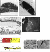
Examples of vascular lesions resulting from deforming stress. (A) Images of the blood–gas barrier (i.e., intraalveolar capillaries) of rats exposed to injurious mechanical ventilation. Note endothelial (A1) and epithelial (A2) blebbing and gaps that are marked by arrows. AS = alveolar space; IE = interstitial edema; PN = polymorphonuclear neutrophil. (Reproduced with permission from Dreyfuss D, et al. Principles and Practices of Mechanical Ventilation. New York: McGraw-Hill, 1994. pp. 793–811.) (B) Images of the blood–gas barrier of rabbits with hydrostatic pulmonary edema. Note blebbing and vesicle formation (B1and B2, thin arrows and asterisks) as well as the large alveolar fenestration with denuded/exposed basement membrane (B3, wide arrow). AE = alveolar edema; BM = basement membrane; End = endothelium. (B1 and B2 reproduced with permission from Reference 54; B3 reproduced with permission from Reference 25.) (C) Images of two adherent endothelial cells (red and yellow) from a frog mesenteric capillary that is exposed to high vascular pressures. Upper panel = en-face view; lower panel = cross-section. Note the intracellular gap formation (G1) and the preserved intercellular tight junction. (Reproduced with permission from Reference 89.) (D) Scanning electron-micrograph of an intraalveolar pulmonary capillary from a mechanically ventilated patient with acute respiratory distress syndrome (D1). Note that the capillary/basement membrane fracture (D2 is magnified view). (Reproduced with permission from Reference 80.)
<b>Figure 2.</b>
Figure 2.
Light microscopic (upper panel) and live tissue images (lower panel) of isolated perfused rat lungs after mechanical ventilation at noninjurious (tidal volume 6 ml/kg) or injurious (tidal volume 40 ml/kg) settings. The perfusate contained propidium iodide, a membrane-impermeant molecule, which on entering the cell emits a red fluorescence when it is interchelated with RNA or DNA. Note the increased cellularity, the perivascular hemorrhage, and the damage to small airway lining cells in the histologic section of the injured lung. Note the prominent red nuclei of transiently or permanently wounded subpleural cells in the live tissue images obtained with laser confocal microscopy. (Reproduced with permission from Reference 95.)
<b>Figure 3.</b>
Figure 3.
Cartoon of putative cellular mechanosensing structures (A) and their response to deforming stress (B). (A) Deformation (strain) of the matrix (basement membrane) generates a force, which is transmitted via adhesion receptors (e.g., integrins) to the cell. To date, over 50 different focal adhesion proteins have been identified that link adhesion receptors to the tension bearing elements of the cytoskeleton (CSK). They are thought to be a major locus of mechanosensing, i.e., they respond to forces that are either generated by the cells (via molecular motors) and are carried via the CSK or which are externally imposed (e.g., in the lung during breathing) and transmitted to the CSK. Moreover, tension-bearing elements of the CSK can connect directly to protein channels (shown in blue), thereby mechanically gating ion flux through them. (B) An externally imposed shape change is associated with the unfolding of excess plasma membrane. As lateral tension of the unfolded plasma membrane increases channel proteins (e.g., mechanosensitive cation channels), that are suspended by hydrophobic matching in the lipid bilayer, undergo a conformational change and ion flux (e.g., Ca2+) increases. The increase in plasma membrane tension triggers a vigorous lipid (and protein) trafficking response (brown and yellow vesicles) that results in a net growth of plasma membrane surface area.
<b>Figure 4.</b>
Figure 4.
Schematic of the cellular response to membrane stress failure. Calcium enters the cell through a plasma membrane defect. Sustained large elevations in intracellular Ca2+ produce necrosis. Smaller transients in intracellular Ca2+ initiate cell repair responses. Cells repair membrane defects but several mechanisms (right-hand side). Mechanism 1 involves lateral flow plasma membrane lipids driven the free energy (analogous to surface tension) at the wound edge. This mechanisms is thought to play a role in the healing of small defects. Mechanism 2 is the fusion of early endosomes with the plasma membrane. Mechanism 3 involves the coalescence of vesicular organelles (usually lysosomes), which form a patch and plugs the wound by Ca2+-induced, site-directed exocytosis. Wounding and repair trigger also the translocation of nuclear transcription factors like NFκ-B, leading to the induction of early stress response genes and thereby initiate proinflammatory signaling cascades.

References

    1. Rubenfeld GD. Epidemiology of acute lung injury. Crit Care Med 2003;31:S276–S284. - PubMed
    1. Moss M, Mannino DM. Race and gender differences in acute respiratory distress syndrome deaths in the United States: An analysis of multiple cause mortality data (1979–1996). Crit Care Med 2002;30:1679–1685. - PubMed
    1. Bersten AD, Edibam C, Hunt T, Moran J. Incidence and mortality of acute lung injury and the acute respiratory distress syndrome in three Australian States. Am J Respir Crit Care Med 2002;165:443–448. - PubMed
    1. Milberg JA, Davis DR, Steinberg KP, Hudson LD. Improved survival of patients with acute respiratory distress syndrome (ARDS): 1983–1993. JAMA 1995;273:306–309. - PubMed
    1. Luhr OR, Antonsen K, Karlsson M, Aardal S, Thorsteinsson A, Frosell CG, Bonde J. Incidence and mortality after acute respiratory failure and acute respiratory distress syndrome in Sweden, Denmark, and Iceland. The ARF Study Group. Am J Respir Crit Care Med 1999;159:1849–1861. - PubMed

Publication types