. 2005 Feb 15;102(7):2649-54.
doi: 10.1073/pnas.0406377102. Epub 2005 Feb 4.

A plasma membrane H+-ATPase is required for the formation of proanthocyanidins in the seed coat endothelium of Arabidopsis thaliana

Affiliations

A plasma membrane H+-ATPase is required for the formation of proanthocyanidins in the seed coat endothelium of Arabidopsis thaliana

Ivan R Baxter et al. Proc Natl Acad Sci U S A. .

Erratum in

  • Proc Natl Acad Sci U S A. 2005 Apr 12;102(15):5635

Abstract

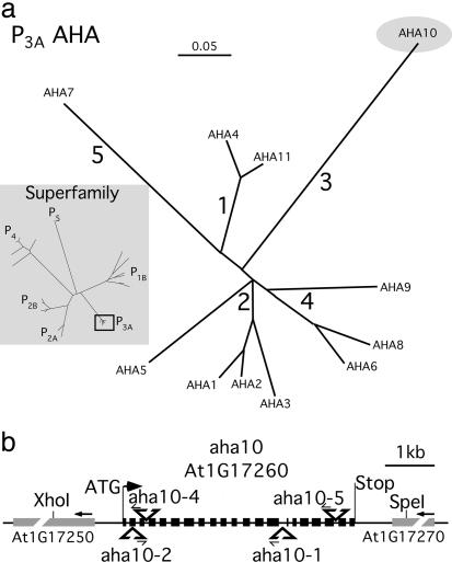
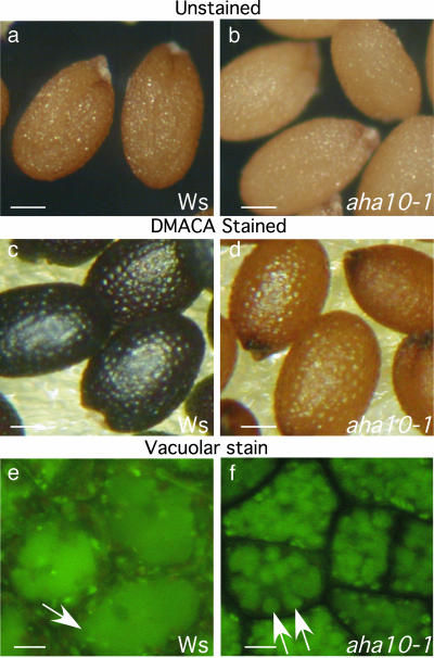
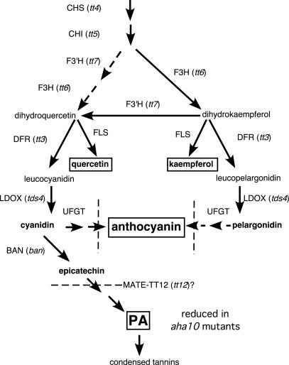
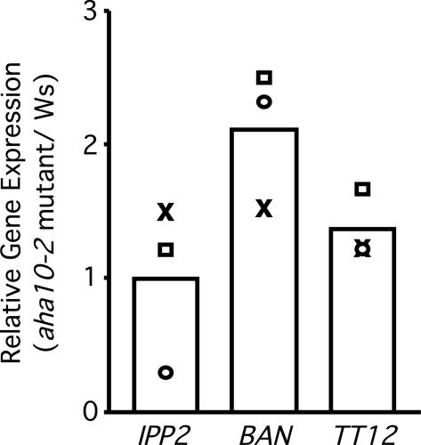
The plasma membrane in plant cells is energized with an electrical potential and proton gradient generated through the action of H+ pumps belonging to the P-type ATPase superfamily. The Arabidopsis genome encodes 11 plasma membrane H+ pumps. Auto-inhibited H+-ATPase isoform 10 (AHA10) is expressed primarily in developing seeds. Here we show that four independent gene disruptions of AHA10 result in seed coats with a transparent testa (tt) phenotype (light-colored seeds). A quantitative analysis of extractable flavonoids in aha10 seeds revealed an approximately 100-fold reduction of proanthocyanidin (PA), one of the two major end-product pigments in the flavonoid biosynthetic pathway. In wild-type seed coat endothelial cells, PA accumulates in a large central vacuole. In aha10 mutants, the formation of this vacuole is impaired, as indicated by the predominance of multiple small vacuoles observed by fluorescence microscopy using a vacuole-specific dye, 5-(and -6)-carboxy 2',7'-dichlorofluorescein diacetate. A similar vacuolar defect was also observed for another tt mutant, tt12, a proton-coupled multidrug and toxic compound extrusion transporter potentially involved in loading provacuoles with a flavonoid intermediate required for PA production. The endothelial cells in aha10 mutants are otherwise healthy, as indicated by the lack of a significant decrease in (i) the accumulation of other flavonoid pathway end products, such as anthocyanins, and (ii) mRNA levels for two endothelium-specific transcripts (TT12 and BAN). Thus, the specific effect of aha10 on vacuolar and PA biogenesis provides genetic evidence to support an unexpected endomembrane function for a member of the plasma membrane H+-ATPase family.


Figures

Fig. 1.
Fig. 1.
The Arabidopsis P-type ATPase superfamily and map of AHA10. (a) Relationship tree showing the autoinhibited H+ P-type ATPase (AHA) family in Arabidopsis. The numbers represent the subfamilies of the plant AHAs identified by Arango et al. (2) and Baxter et al. (3). (Inset) Relationship tree showing the subfamilies of plant P-type ATPases identified by Baxter et al. (3). A box surrounds the AHA branch shown in the main part of the figure. (b) Sites of T-DNA insertions in aha10 mutants. Exons are shown as black boxes; the coding regions of the two adjacent genes are shown as gray boxes. Arrowheads indicate the direction of transcription. T-DNA insertion points are indicated by triangles, and the left border sequence is shown with arrows. The left border sequences of aha10–1 (3313ATGAAAAATCAcatcccggacgatatattgt) and aha10–2 (tgactgacagACTTGAAGAG266) (AHA10 sequence in capital letters, numbering from ATG) were determined by sequencing of PCR-amplified borders. The border sequences of aha10-4 and aha10-5 can be found at the SIGnAL web site (http://signal.salk.edu/cgi-bin/tdnaexpress). The restriction sites used to create the complementation clone are shown.
Fig. 2.
Fig. 2.
AHA10 loss of function results in a tt phenotype with reduced PA and altered vacuoles. (a and b) Light microscope images of wild-type (a) and aha10 (b) seeds. (c and d) DMACA staining of Ws and aha10-1 seeds. DMACA changes color when it binds to 3′,4′ flavon diols, including PA and its precursors. (e and f) Seed coat endothelial cells in aha10 mutants display a vacuolar biogenesis defect. Developing seeds were dissected and stained with the vacuole-specific dye 5-(and -6)-carboxy 2′,7′-dichlorofluorescein diacetate. Arrows in e and f point to the large and small vacuoles, respectively. (Scale bars: a–d, 0.1 mm; e and f, 10 μm.)
Fig. 3.
Fig. 3.
Flavonoid pathway in Arabidopsis. Pathway diagram is derived from ref. . The known elements of the flavonoid pathway are indicated. The protein names are capitalized; the corresponding mutant line in Arabidopsis is in italics. The different chemical species are indicated in lowercase. Bold letters indicate flavonoids quantified in Table 1. End products are boxed. Dotted line perpendicular to arrow indicates a hypothetical membrane transport step. Plants with disruptions in transcription factor genes TT1, TT2, TT8, TT16, TTG1, and TTG2 have been shown to have tt phenotypes, as well. tds, tannin deficient seed; CHS, chalcone synthase; CHI, chalcone isomerase; F3H, flavone 3 hydroxylase; F3′H, flavone 3′ hydroxylase; DFR, dihydroflavonol reductase; FLS, flavonol synthase; UFGT, UDP glucose-flavonoid 3-O-glucosyl transferase.
Fig. 4.
Fig. 4.
Expression levels are not decreased for two PA pathway-specific genes. Quantitative RT-PCR of BAN and TT12 transcripts in green siliques. The levels of BAN, TT12, and a control gene (IPP2) were compared with the APX3 gene for each RNA sample. In controls, the primers for TT12 and BAN were shown to specifically amplify their respective cDNAs from siliques at a level >1,000 times that found by using template RNA from 2-week-old seedling tissue. The relative levels of gene expression are shown as the ratio of mutant to wild type for each transcript. The bars represent the average of three independent trials, which are shown individually as open circles, open squares, and “X”s.

Similar articles

Cited by

References

    1. Sze, H., Li, X. & Palmgren, M. G. (1999) Plant Cell 11, 677-690. - PMC - PubMed
    1. Arango, M., Gévaudant, F., Oufattole, M. & Boutry, M. (2003) Planta 216, 355-365. - PubMed
    1. Baxter, I., Tchieu, J., Sussman, M. R., Boutry, M., Palmgren, M. G., Gribskov, M., Harper, J. F. & Axelsen, K. B. (2003) Plant Physiol. 132, 618-628. - PMC - PubMed
    1. Harper, J. F., Manney, L. & Sussman, M. R. (1994) Mol. Gen. Genet. 244, 572-587. - PubMed
    1. Debeaujon, I., Nesi, N., Perez, P., Devic, M., Grandjean, O., Caboche, M. & Lepiniec, L. (2003) Plant Cell 15, 2514-2531. - PMC - PubMed

Publication types

MeSH terms