. 2005 Mar;115(3):733-8.
doi: 10.1172/JCI23001.

Fluid shear stress inhibits vascular inflammation by decreasing thioredoxin-interacting protein in endothelial cells

Affiliations

Fluid shear stress inhibits vascular inflammation by decreasing thioredoxin-interacting protein in endothelial cells

Hideyuki Yamawaki et al. J Clin Invest. 2005 Mar.

Abstract

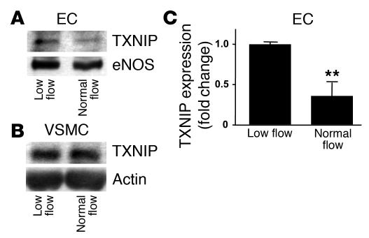
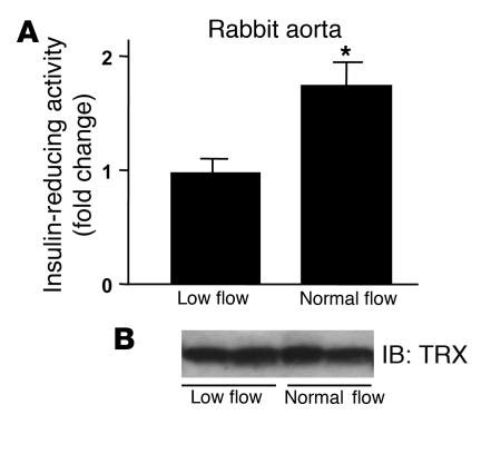
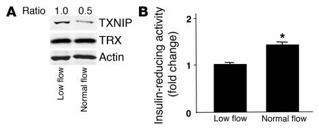
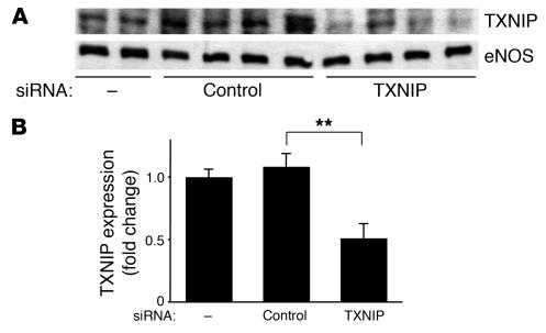
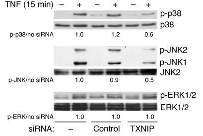
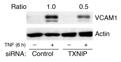
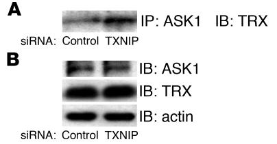
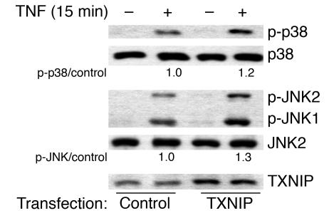
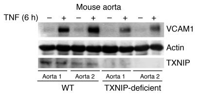
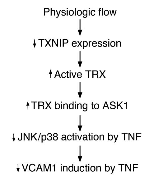
Regions in the vasculature that are exposed to steady laminar blood flow are protected from atherosclerosis as compared with regions where flow is disturbed. We found that flow decreased TNF-mediated VCAM1 expression by inhibiting JNK and p38. JNK inhibition correlated with inhibition of apoptosis signal-regulating kinase 1 (ASK1), a JNK and p38 activator. Thioredoxin-interacting protein (TXNIP) is a stress-responsive protein that inhibits thioredoxin (TRX) activity. Since thioredoxin inhibits ASK1, we hypothesized that changes in TXNIP-TRX-ASK1 interactions mediate the antiinflammatory effects of flow. To explore this, we used perfused vessels and cultured ECs. Exposure of rabbit aortae or ECs to normal flow (12 dyn/cm2, 24 hours) was associated with decreased TXNIP expression and increased TRX activity compared with exposure to low flow (0.4 dyn/cm2). Normal flow inhibited TNF activation of JNK/p38 and VCAM1 expression. In cultured ECs, reduction of TXNIP expression by small interfering RNA increased TRX binding to ASK1 and inhibited TNF activation of JNK/p38 and VCAM1 expression. Conversely, overexpression of TXNIP stimulated JNK and p38. In aortae from TXNIP-deficient mice, TNF-induced VCAM1 expression was inhibited. The data suggest that TXNIP and TRX are key components of biomechanical signal transduction and establish them as potentially novel regulators of TNF signaling and inflammation in ECs.


Figures

Figure 1
Figure 1
Normal flow downregulated TXNIP expression in ECs. After rabbit aortae were exposed to low flow (0.4 dyn/cm2) or normal flow (12 dyn/cm2) for 24 hours, EC (A) and VSMC (B) proteins were selectively purified, and immunoblotting was performed (n = 4–5). Equal protein loading was confirmed with eNOS or actin antibody. (C) TXNIP expression is shown as fold change relative to low flow. **P < 0.01 vs. low flow.
Figure 2
Figure 2
Normal flow increased the activity but not the expression of TRX in ECs. After rabbit aortae were exposed to low flow (0.4 dyn/cm2) or normal flow (12 dyn/cm2) for 24 hours, EC lysates were harvested. (A) TRX activity was determined by the insulin-reducing assay. Results are shown as fold change relative to low flow (n = 4). *P < 0.05 vs. low flow. (B) TRX expression was unchanged in low and normal flow (n = 5).
Figure 3
Figure 3
Normal flow downregulated TXNIP expression and upregulated TRX activity in HUVECs. After HUVECs were exposed to low flow (0.4 dyn/cm2) or normal flow (12 dyn/cm2) for 24 hours, proteins were harvested. (A) TXNIP and TRX expression was determined by immunoblotting (n = 4). Equal protein loading was confirmed with actin antibody. TXNIP expression is shown as fold change relative to low flow. The ratio in normal flow differed significantly from that in low flow (n = 4, P < 0.05). (B) TRX activity was determined by the insulin-reducing assay. Results are shown as fold change relative to low flow (n = 4). *P < 0.05 vs. low flow.
Figure 4
Figure 4
TXNIP protein expression was decreased by siRNA in HUVECs. HUVECs were transfected with either control or TXNIP siRNA. (A) TXNIP expression was determined by immunoblotting from 4–6 independent experiments. Equal protein loading was confirmed with eNOS antibody. (B) TXNIP expression is shown as fold change relative to no siRNA. **P < 0.01 vs. control siRNA.
Figure 5
Figure 5
TXNIP siRNA inhibited TNF activation of p38 and JNK but not ERK1/2 in HUVECs. After HUVECs were transfected with either control or TXNIP siRNA, TNF (10 ng/ml) was added for 15 minutes. MAPK activation was determined by immunoblotting using phospho-specific (p-) antibody. Equal loading was confirmed with total MAPK antibodies. Representative blots from 3 independent experiments are shown. Quantitation of the ratio of phospho-MAPK in lysates from cells treated with control or TXNIP siRNA to phospho-MAPK in lysates from cells not treated with siRNA is shown below each panel (average of 3 determinations).
Figure 6
Figure 6
TXNIP siRNA inhibited TNF-induced VCAM1 expression in HUVECs. After HUVECs were transfected with either control or TXNIP siRNA, TNF-α (10 ng/ml) was added for 6 hours. VCAM1 expression was determined by immunoblotting from 3 independent experiments. Equal protein loading was confirmed with actin antibody. VCAM1 expression is shown as fold change relative to control siRNA.
Figure 7
Figure 7
TXNIP siRNA increased TRX binding to ASK1 in HUVECs. (A) Interaction of ASK1 with TRX was examined by immunoblotting with TRX antibody after HUVEC lysates were immunoprecipitated with ASK1 antibody. (B) Equal loading was confirmed with ASK1, TRX, and actin antibodies. Results are representative of 4 independent experiments.
Figure 8
Figure 8
TXNIP overexpression augmented TNF-induced p38 and JNK activation in BAECs. BAECs were transfected with either control (pcDNA3.1) or pcDNA3.1-TXNIP and then treated with TNF-α (10 ng/ml) for 15 minutes. Activation of p38 and JNK was determined by immunoblotting using phospho-specific antibody. Equal loading was confirmed with total MAPK antibodies. Representative blots from 3–4 independent experiments are shown.
Figure 9
Figure 9
TNF-induced VCAM1 expression was decreased in TXNIP-deficient mouse aorta. After aortae from HcB-19 and control C3H mice were treated with TNF (15 ng/ml, 6 hours), vessel protein was harvested. Expression of VCAM1 and TXNIP was determined by immunoblotting in 2 aortic samples from 2 animals of each strain. Equal protein loading was confirmed with actin antibody.
Figure 10
Figure 10
Flow regulates TXNIP in ECs. Chronic exposure to normal flow decreases TXNIP expression, and this results in increased TRX binding to ASK1. This inhibits cytokine activation of the JNK-p38 pathway and prevents proinflammatory events such as VCAM1 expression.

References

    1. Gimbrone MA, Jr, Topper JN, Nagel T, Anderson KR, Garcia-Cardena G. Endothelial dysfunction, hemodynamic forces, and atherogenesis. Ann. N. Y. Acad. Sci. 2000;902:230–239. - PubMed
    1. Traub O, Berk BC. Laminar shear stress: mechanisms by which endothelial cells transduce an atheroprotective force. Arterioscler. Thromb. Vasc. Biol. 1998;18:677–685. - PubMed
    1. Ross R. Atherosclerosis: an inflammatory disease. N. Engl. J. Med. 1999;340:115–126. - PubMed
    1. Surapisitchat J, et al. Fluid shear stress inhibits TNF-alpha activation of JNK but not ERK1/2 or p38 in human umbilical vein endothelial cells: inhibitory crosstalk among MAPK family members. Proc. Natl. Acad. Sci. U. S. A. 2001;98:6476–6481. - PMC - PubMed
    1. Liu Y, Yin G, Surapisitchat J, Berk BC, Min W. Laminar flow inhibits TNF-induced ASK1 activation by preventing dissociation of ASK1 from its inhibitor 14-3-3. J. Clin. Invest. 2001;107:917–923. - PMC - PubMed

Publication types

MeSH terms

Substances