. 2005 Feb 15;174(4):1962-70.
doi: 10.4049/jimmunol.174.4.1962.

CCR8 is expressed by antigen-elicited, IL-10-producing CD4+CD25+ T cells, which regulate Th2-mediated granuloma formation in mice

Affiliations

CCR8 is expressed by antigen-elicited, IL-10-producing CD4+CD25+ T cells, which regulate Th2-mediated granuloma formation in mice

Christine M Freeman et al. J Immunol. .

Abstract

CCR8 was initially described as a Th2 cell-restricted receptor, but this has not been fully tested in vivo. The present study used ex vivo and in vivo approaches to examine the distribution and functional significance of CCR8 among CD4+ T cells. Populations of cytokine-secreting CD4+ T cells were generated in primed mice with Th1 or Th2 cell-mediated pulmonary granulomas, respectively elicited by i.v. challenge with either Mycobacteria bovis purified protein derivative- or Schistosoma mansoni egg Ag (SEA)-coated beads. Cytokine-producing CD4+ T cells were isolated from Ag-stimulated draining lymph node cultures by positive selection. Quantitative analysis of cytokine mRNA indicated enriched populations of IFN-gamma-, IL-4-, and IL-10-producing cells. Analysis of chemokine receptor mRNA indicated that IL-10+ cells selectively expressed CCR8 in the SEA bead-elicited type 2 response. The IL-10+CCR8+ populations were CD25+ and CD44+ but lacked enhanced Foxp3 expression. Adoptive transfer to naive recipients indicated that IL-10+ T cells alone could not transfer type 2 inflammation. Analysis of SEA bead-challenged CCR8-/- mice indicated significantly impaired IL-10 production as well as reductions in granuloma eosinophils. Adoptive transfer of CD4+CCR8+/+ T cells corrected cytokine and inflammation defects, but the granuloma eosinophil recruitment defect persisted when donor cells were depleted of IL-10+ cells. Accordingly, local IL-10 production correlated with CCR8 ligand (CCL1) expression and the appearance of CCR8+ cells in granulomatous lungs. Thus, IL-10-producing, CCR8+CD4+CD25+CD44+ T cells are generated during SEA challenge, which augment the Th2-mediated eosinophil-rich response to the parasite Ags.


Figures

FIGURE 1
FIGURE 1
Isolation of IFN-γ, IL-4, and IL-10 cytokine-producing CD4+ T cells from draining lymph nodes during type 1 (PPD) and type 2 (SEA) granuloma formation. Secondary lung granulomas were induced in CBA/J mice as described in Materials and Methods. On day 3, draining lymph nodes were collected and cultured overnight with Ag. CD4+ cytokine-secreting populations were isolated using MACS. The CD4+ nonenriched population serves as a control. Relative mRNA transcript levels were measured for IFN-γ (A), IL-4 (B), and IL-10 (C) using real-time PCR. Type 1 (PPD) response (▪); type 2 (SEA) response (▨). Bars are mean arbitrary units ± SD and are derived from three separate experiments.
FIGURE 2
FIGURE 2
Chemokine receptor transcript association with IL-10-, IL-4-, and IFN-γ-secreting CD4+ T cells isolated from draining lymph nodes of CBA/J mice undergoing type 1 or type 2 granulomatous responses. Transcript levels of the chemokine receptors CCR3 (A), CCR4 (B), CCR5 (C), CCR6 (D), CCR7 (E), and CCR8 (F) were measured by real-time PCR. Type 1 (PPD) response (▪); type 2 (SEA) response (▨). E, Dashed line represents transcript levels in naive CD4+ T cells. Bars are mean arbitrary units ± SD and are derived from three separate experiments.
FIGURE 3
FIGURE 3
Chemokine receptor and cytokine transcript association with CD44+ and CD25+ activated CD4+ T cells isolated from draining lymph nodes of CBA/J mice during type 2 granuloma formation. Following column depletion of either CD44+ or CD25+ cells, CD4+ T cell mRNA transcripts were measured by real-time PCR. Bars are mean arbitrary units ± SD and are derived from two separate experiments. *, p < 0.05 compared with the total CD4+ population.
FIGURE 4
FIGURE 4
Foxp3 transcript expression by CD4+ T cells and CD4+ subpopulations isolated from the draining lymph nodes of mice undergoing type 2 granuloma formation. After overnight culture with Ag, CD4+ T cells were either enriched for IL-10 or depleted of CD25 and IL-10. Foxp3 mRNA transcript levels were measured by real-time PCR. Bars are mean arbitrary units ± SD and are derived from two separate experiments. *, p < 0.05 compared with the CD4+ control.
FIGURE 5
FIGURE 5
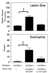
Enriched CD4+ IL-10-secreting T cells fail to transfer anamnestic type 2 granulomatous responses to naive mice. Draining lymph nodes were collected from SEA-sensitized and -challenged wild-type (sCCR8+/+) donors and then cultured overnight with Ag. CD4+ and CD4+IL-10+ enriched cells were isolated from these cultures as described in Materials and Methods, and then 1 × 10⁶ cells were adoptively transferred i.v. into naive wild-type (nCCR8+/+) syngeneic recipients. Recipient mice were challenged with SEA beads the following day. Granulomas were examined on day 4 after bead challenge. Top panel, Granuloma cross-sectional area; bottom panel, eosinophil content of dispersed granulomas. Data are representative of two separate experiments. Bars are means ± SEM. * p < 0.05 compared with the mice that did not receive cells.
FIGURE 6
FIGURE 6
CCR8 gene knockout is associated with impaired IL-10 production by draining lymph node cultures during primary and secondary type 2 (SEA) granulomatous responses. Primary and secondary lung granulomas were induced in CCR8+/+ and CCR8−/− mice as described in Materials and Methods. On day 4, draining lymph nodes were collected and cultured with Ag for 24 or 48 h for secondary or primary responses, respectively. Cytokine levels were determined by ELISA. Bars are means ± SEM of Ag-elicited cytokine levels from a representative experiment. Left sides of panels are primary SEA responses; right sides of panels are secondary SEA responses. CCR8+/+ mice (▪); CCR8−/− mice (▨). * p < 0.05 comparing CCR8+/+ to CCR8−/− responses.
FIGURE 7
FIGURE 7
Wild-type CCR8+/+CD4+ T cells from SEA-sensitized CCR8 +/+ mice reconstitute type 2 granulomatous responses of CCR8−/− mice. Donor mice were sensitized with 3000 S. mansoni eggs i.p. on days 1 and 10. On day 13, spleens were collected and cultured overnight with Ag. CD4+ T cells were isolated using negative selection. A total of 1 × 10⁶ cells was i.v. transferred to SEA-sensitized CCR8−/− mice. Recipients were challenged with SEA beads the following day. Granulomas were examined on day 4 after bead challenge. Top panel, Granuloma cross-sectional area; bottom panel, eosinophil content of dispersed granulomas. Data are representative of three separate experiments. Bars are means ± SEM.
FIGURE 8
FIGURE 8
Wild-type CCR8+CD4+ T cells depleted of IL-10-secreting cells fail to reconstitute type 2 granulomatous responses of CCR8−/− mice. Donor mice were given 3000 S. mansoni eggs i.p. on days 1 and 10. On day 13, spleens were collected and cultured overnight with Ag. CD4+ T cells were isolated using negative selection. IL-4- or IL-10-secreting populations were depleted as described in Materials and Methods. A total of 1 × 10⁶ cells was i.v. administered to SEA-sensitized CCR8−/− mice. Recipients were challenged with SEA beads on the following day. Granulomas were examined on day 4 after bead challenge. Top panel, Granuloma cross-sectional area; bottom panel, eosinophil content of dispersed granulomas. Data are representative of three separate experiments. Bars are means ± SEM.
FIGURE 9
FIGURE 9
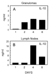
IL-10 expression in granulomatous lungs and draining lymph nodes during secondary type 2 (SEA) granulomatous responses. Groups of S. mansoni egg-sensitized CBA/J mice were challenged with SEA beads, and then on days 1, 2, 4, and 8, intact granulomas (1000/ml) and dispersed draining lymph nodes (5 3 106 cells/ml) were cultured, respectively, for 48 and 24 h with 5 μg/ml SEA. IL-10 levels were determined in culture supernatants by ELISA. A, Granuloma cultures; B, lymph node cultures. Three to four mice per point.
FIGURE 10
FIGURE 10
CCL1 (TCA-3) (A) and CCR8 (B) transcript induction in lungs and draining lymph nodes during secondary type 2 (SEA) granulomatous responses. Type 2 lung granulomas were induced in groups of S. mansoni egg-sensitized CBA/J mice as described in Materials and Methods. Lungs and draining lymph nodes were collected before bead challenge (day 0) and on days 1, 2, 3, 4, and 8 post-bead challenge. The freshly isolated tissues were used for transcript analysis by real-time PCR. Data are derived from two separate experiments for a total of five to six mice per point.
FIGURE 11
FIGURE 11
CCL1 and IL-10 transcript expression in lungs of CCR8+/+ wild-type and CCR8−/− mice during secondary type 2 (SEA) granulomatous responses. Type 2 lung granulomas were induced in groups of S. mansoni egg-sensitized CCR8+/+ or CCR8−/− mice as described in Materials and Methods. Lungs were collected on day 3 post-SEA bead challenge. The freshly isolated tissue was used for transcript analysis by real-time PCR. A, CCL1 transcript levels; B, IL-10 transcript levels. Data are derived from four to five individual mice per point.*, p < 0.05 comparing CCR8+/+ to CCR8−/− levels.

Similar articles

Cited by

References

    1. Thelen M. Dancing to the tune of chemokines. Nat Immunol. 2001;2:129. - PubMed
    1. Qin S, Rottman JB, Myers P, Kassam N, Weinblatt M, Loetscher M, Koch AE, Moser B, Mackay CR. The chemokine receptors CXCR3 and CCR5 mark subsets of T cells associated with certain inflammatory reactions. J Clin Invest. 1998;101:746. - PMC - PubMed
    1. Bonecchi R, Bianchi G, Bordignon PP, D’Ambrosio D, Lang R, Borsatti A, Sozzani S, Allavena P, Gray PA, Mantovani A, Sinigaglia F. Differential expression of chemokine receptors and chemotactic responsiveness of type 1 T helper cells (Th1s) and Th2s. J Exp Med. 1998;187:129. - PMC - PubMed
    1. Loetscher P, Uguccioni M, Bordoli L, Baggiolini M, Moser B, Chizzolini C, Dayer JM. CCR5 is characteristic of Th1 lymphocytes. Nature. 1998;391:344. - PubMed
    1. Sallusto F, Mackay CR, Lanzavecchia A. Selective expression of the eotaxin receptor CCR3 by human T helper 2 cells. Science. 1997;277:2005. - PubMed

Publication types

MeSH terms