Electrostatic modeling predicts the activities of orthopoxvirus complement control proteins
- PMID: 15699145
- PMCID: PMC4138803
- DOI: 10.4049/jimmunol.174.4.2143
Electrostatic modeling predicts the activities of orthopoxvirus complement control proteins
Abstract
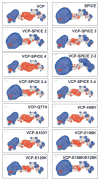
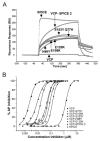
Regulation of complement activation by pathogens and the host are critical for survival. Using two highly related orthopoxvirus proteins, the vaccinia and variola (smallpox) virus complement control proteins, which differ by only 11 aa, but differ 1000-fold in their ability to regulate complement activation, we investigated the role of electrostatic potential in predicting functional activity. Electrostatic modeling of the two proteins predicted that altering the vaccinia virus protein to contain the amino acids present in the second short consensus repeat domain of the smallpox protein would result in a vaccinia virus protein with increased complement regulatory activity. Mutagenesis of the vaccinia virus protein confirmed that changing the electrostatic potential of specific regions of the molecule influences its activity and identifies critical residues that result in enhanced function as measured by binding to C3b, inhibition of the alternative pathway of complement activation, and cofactor activity. In addition, we also demonstrate that despite the enhanced activity of the variola virus protein, its cofactor activity in the factor I-mediated degradation of C3b does not result in the cleavage of the alpha' chain of C3b between residues 954-955. Our data have important implications in our understanding of how regulators of complement activation interact with complement, the regulation of the innate immune system, and the rational design of potent complement inhibitors that might be used as therapeutic agents.
Figures


Similar articles
-
Evolutionary history of orthopoxvirus proteins similar to human complement regulators.Gene. 2005 Aug 1;355:40-7. doi: 10.1016/j.gene.2005.05.008. Gene. 2005. PMID: 16023794 Free PMC article.
-
Species Specificity of Vaccinia Virus Complement Control Protein for the Bovine Classical Pathway Is Governed Primarily by Direct Interaction of Its Acidic Residues with Factor I.J Virol. 2017 Sep 12;91(19):e00668-17. doi: 10.1128/JVI.00668-17. Print 2017 Oct 1. J Virol. 2017. PMID: 28724763 Free PMC article.
-
Interaction of vaccinia virus complement control protein with human complement proteins: factor I-mediated degradation of C3b to iC3b1 inactivates the alternative complement pathway.J Immunol. 1998 Jun 1;160(11):5596-604. J Immunol. 1998. PMID: 9605165
-
Variola virus immune evasion proteins.Microbes Infect. 2003 Sep;5(11):1049-56. doi: 10.1016/s1286-4579(03)00194-1. Microbes Infect. 2003. PMID: 12941397 Review.
-
Equination (inoculation of horsepox): An early alternative to vaccination (inoculation of cowpox) and the potential role of horsepox virus in the origin of the smallpox vaccine.Vaccine. 2017 Dec 19;35(52):7222-7230. doi: 10.1016/j.vaccine.2017.11.003. Epub 2017 Nov 11. Vaccine. 2017. PMID: 29137821 Review.
Cited by
-
Activator-specific requirement of properdin in the initiation and amplification of the alternative pathway complement.Blood. 2008 Jan 15;111(2):732-40. doi: 10.1182/blood-2007-05-089821. Epub 2007 Oct 4. Blood. 2008. PMID: 17916747 Free PMC article.
-
Insight into mode-of-action and structural determinants of the compstatin family of clinical complement inhibitors.Nat Commun. 2022 Sep 20;13(1):5519. doi: 10.1038/s41467-022-33003-7. Nat Commun. 2022. PMID: 36127336 Free PMC article.
-
Complement evasion by human pathogens.Nat Rev Microbiol. 2008 Feb;6(2):132-42. doi: 10.1038/nrmicro1824. Nat Rev Microbiol. 2008. PMID: 18197169 Free PMC article. Review.
-
Identification of hot spots in the variola virus complement inhibitor (SPICE) for human complement regulation.J Virol. 2008 Apr;82(7):3283-94. doi: 10.1128/JVI.01935-07. Epub 2008 Jan 23. J Virol. 2008. PMID: 18216095 Free PMC article.
-
Complement Evasion Strategies of Viruses: An Overview.Front Microbiol. 2017 Jun 16;8:1117. doi: 10.3389/fmicb.2017.01117. eCollection 2017. Front Microbiol. 2017. PMID: 28670306 Free PMC article. Review.
References
-
- Liszewski MK, Atkinson JP. Regulatory proteins of complement. In: Volanakis JE, Frank MM, editors. The Human Complement System in Health and Disease. Decker; New York: 1998. p. 149.
-
- Hourcade D, Liszewski MK, Krych-Goldberg M, Atkinson JP. Functional domains, structural variations and pathogen interactions of MCP, DAF and CR1. Immunopharmacology. 2000;49(103) - PubMed
-
- Herbert A, O'Leary J, Krych-Goldberg M, Atkinson JP, Barlow PN. Three-dimensional structure and flexibility of proteins of the RCA family—a progress report. Biochem Soc Trans. 2002;30:990. - PubMed
-
- Mastellos D, Morikis D, Isaacs SN, Holland MC, Strey CW, Lambris JD. Complement: structure, functions, evolution, and viral molecular mimicry. Immunol Res. 2003;27:367. - PubMed
-
- Mullick J, Kadam A, Sahu A. Herpes and pox viral complement control proteins: “the mask of self”. Trends Immunol. 2003;24:24. - PubMed
Publication types
MeSH terms
Substances
Grants and funding
LinkOut - more resources
Full Text Sources
Other Literature Sources

