. 2005 Feb 14;168(4):567-73.
doi: 10.1083/jcb.200405120. Epub 2005 Feb 7.

Live imaging of wound inflammation in Drosophila embryos reveals key roles for small GTPases during in vivo cell migration

Affiliations

Live imaging of wound inflammation in Drosophila embryos reveals key roles for small GTPases during in vivo cell migration

Brian Stramer et al. J Cell Biol. .

Abstract

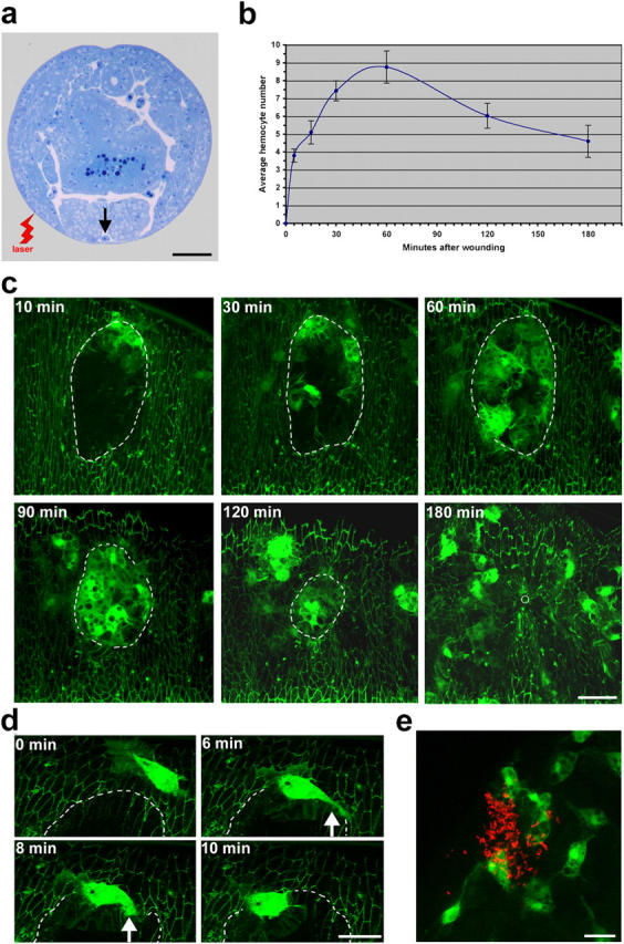
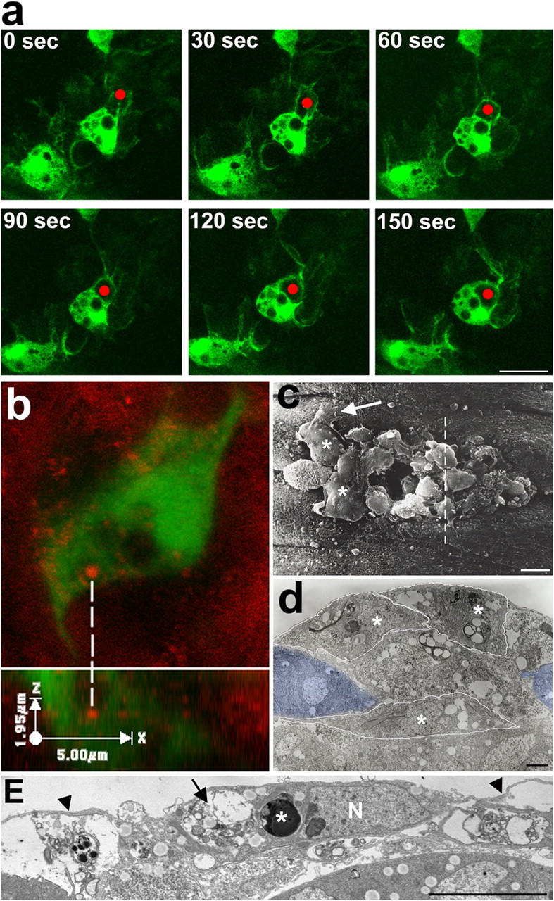
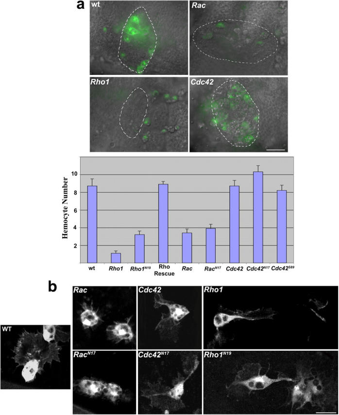
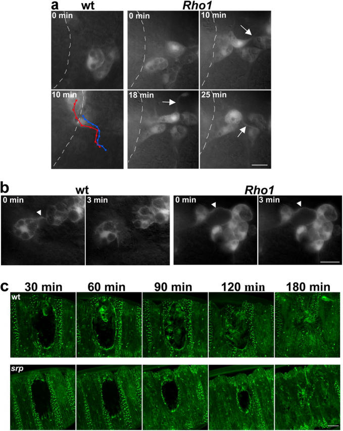
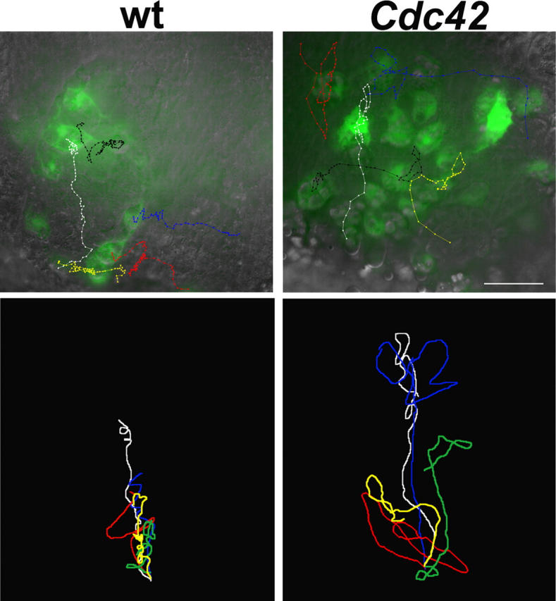
Aa robust inflammatory response to tissue damage and infection is conserved across almost all animal phyla. Neutrophils and macrophages, or their equivalents, are drawn to the wound site where they engulf cell and matrix debris and release signals that direct components of the repair process. This orchestrated cell migration is clinically important, and yet, to date, leukocyte chemotaxis has largely been studied in vitro. Here, we describe a genetically tractable in vivo wound model of inflammation in the Drosophila melanogaster embryo that is amenable to cinemicroscopy. For the first time, we are able to examine the roles of Rho-family small GTPases during inflammation in vivo and show that Rac-mediated lamellae are essential for hemocyte motility and Rho signaling is necessary for cells to retract from sites of matrix- and cell-cell contacts. Cdc42 is necessary for maintaining cellular polarity and yet, despite in vitro evidence, is dispensable for sensing and crawling toward wound cues.


Figures

Figure 1.
Figure 1.
Hemocyte recruitment to laser wounds in the embryo. (a) Transverse resin section through a Drosophila embryo at stage 15 to indicate the site of laser wounding relative to location of ventral hemocytes (arrow). (b) Graph illustrating the temporal recruitment of hemocytes to the wound. (c) Stills showing hemocyte recruitment during the 3-h repair period after wounding a fly embryo expressing both Pxn-GFP and E-cadherin-GFP to reveal hemocytes and epithelium, respectively. (d) High magnification detail of a hemocyte with large polarized lamellar ruffles as it makes a typical “U” turn toward the wound and retracts its tail (arrow) in the process. (e) Pxn-GFP–expressing hemocyte recruitment 1 h after creating a septic wound by delivery of RFP-tagged E. coli with a tungsten needle. Wound edges indicated with dashed lines. Bars: (a) 50 μm; (c) 20 μm; (d and e) 10 μm.
Figure 2.
Figure 2.
Phagocytic engulfment of wound debris by hemocytes at the wound site. (a) Stills from a movie of a Pxn-GFP–expressing embryo, capturing a hemocyte in the act of engulfing wound debris (red dot). (b) Confocal image of red-labeled epithelial debris within a phagocytic vacuole of a Pxn-GFP–expressing hemocyte. Bottom panel represents the “Z” series reconstruction. (c) Scanning electron microscopic view of a 1-h wound showing cell debris and hemocytes (asterisks) with spreading lamellae (arrow). (d) Transmission electron microscopic view of a wound corresponding to the dotted line in panel c and revealing several large hemocytes (outlined by white lines; nuclei indicated with asterisks), swollen with vacuoles containing cell debris. Epidermal wound edge cells are shaded blue. (e) Detail from an adjacent transmission electron microscopic view showing a hemocyte with large lamellae (arrowheads) and containing both apoptotic corpses (asterisk) and vacuoles filled with necrotic debris (arrow). N, cell nucleus. Bars: (a) 10 μm; (c) 5 μm; (d) 1 μm; (e) 5 μm.
Figure 3.
Figure 3.
Hemocyte recruitment after wounding embryos mutant for small GTPases. (a) Dual GFP/DIC images taken 1 h after wounding Rac, Rho, or Cdc42 mutant embryos expressing GFP in hemocytes. Graphic representation of hemocyte numbers recruited to wounds in each genotype. (b) High magnification details of typical hemocyte morphologies. Wild-type hemocytes have broad lamellae, which are much reduced in Rac mutants and in hemocytes expressing the Rac N17 dominant-negative transgene. Cdc42 mutants and dominant-negative Cdc42 N17-expressing hemocytes frequently exhibit bi- and tripolar lamellae. In Rho1 mutants, and Rho1 N19 dominant-negative–expressing hemocytes, cells are often stretched out and occasionally leave remnants of their tail ends behind as they attempt to migrate forward. Bars: (a) 20 μm; (b) 10 μm.
Figure 4.
Figure 4.
Rho1 mutant hemocytes are incapable of retracting cell–cell and cell–matrix contacts. (a) Stills from a time-lapse movie revealing how hemocytes in Rho1 mutant embryos have difficulty retracting their tails (arrows). Eventually, the cell snaps itself forward only to be held by another tether. In contrast, over the same time course, hemocytes from wild-type embryos show rapid directed motility toward the wound. (b) Hemocytes in Rho1 mutant embryos also exhibit persistent links to one another (right, arrowheads), whereas in wild-type hemocytes cell–cell contacts (left, arrowhead) are transient. (c) Comparison of the rate of reepithelialization in sGMCA embryos (revealing both hemocytes and epithelium), which are otherwise wild type or mutant for serpent and thus missing hemocytes. Bars, 10 μm.
Figure 5.
Figure 5.
Abnormal wound migration in Cdc42 mutant hemocytes. Movies from Pxn-GFP, crq-GFP embryos have been used to track the pathways of hemocytes within wounds in wild type versus Cdc42 mutants; tracks have been superimposed on the final still from each of these movies. Bottom panels illustrate the track of five hemocytes each within the wound zone of wild type and Cdc42 mutants; the tracks have all been initiated from the same point and illustrate how much more active mutant hemocytes are than their wild-type counterparts. Bar, 20 μm.

References

    1. Allen, W.E., D. Zicha, A.J. Ridley, and G.E. Jones. 1998. A role for Cdc42 in macrophage chemotaxis. J. Cell Biol. 141:1147–1157. - PMC - PubMed
    1. Cho, N.K., L. Keyes, E. Johnson, J. Heller, L. Ryner, F. Karim, and M.A. Krasnow. 2002. Developmental control of blood cell migration by the Drosophila VEGF pathway. Cell. 108:865–876. - PubMed
    1. Chou, J., N.A. Burke, A. Iwabu, S.C. Watkins, and A. Wells. 2003. Directional motility induced by epidermal growth factor requires Cdc42. Exp. Cell Res. 287:47–56. - PubMed
    1. Cox, D., P. Chang, Q. Zhang, P.G. Reddy, G.M. Bokoch, and S. Greenberg. 1997. Requirements for both Rac1 and Cdc42 in membrane ruffling and phagocytosis in leukocytes. J. Exp. Med. 186:1487–1494. - PMC - PubMed
    1. Dovi, J.V., L.K. He, and L.A. DiPietro. 2003. Accelerated wound closure in neutrophil-depleted mice. J. Leukoc. Biol. 73:448–455. - PubMed

Publication types

MeSH terms

Substances