. 2005 Feb 22;102(8):2886-91.
doi: 10.1073/pnas.0409872102. Epub 2005 Feb 7.

Characterization of CD56-/CD16+ natural killer (NK) cells: a highly dysfunctional NK subset expanded in HIV-infected viremic individuals

Affiliations

Characterization of CD56-/CD16+ natural killer (NK) cells: a highly dysfunctional NK subset expanded in HIV-infected viremic individuals

Domenico Mavilio et al. Proc Natl Acad Sci U S A. .

Abstract

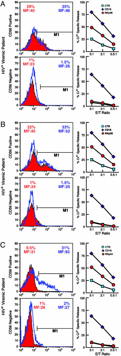
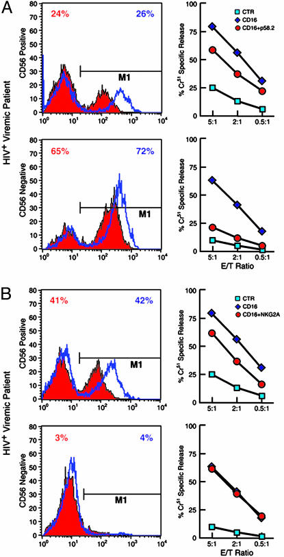
Natural killer (NK) cells are an important component of the innate immune response against viral infections. NK cell-mediated cytolytic activity is defective in HIV-infected individuals with high levels of viral replication. In the present study, we examined the phenotypic and functional characteristics of an unusual CD56(-)/CD16(+) (CD56(-)) NK subset that is greatly expanded in HIV-viremic individuals. The higher level of expression of inhibitory NK receptors and the lower level of expression of natural cytotoxicity receptors observed in the CD56(-) NK fraction compared with that of CD56(+) NK cells was associated with extremely poor in vitro cytotoxic function of this subset. In addition, the secretion of certain cytokines known to be important in initiating antiviral immune responses was markedly reduced in the CD56(-), as compared with the CD56(+) NK cell subset. These data suggest that the expansion of this highly dysfunctional CD56(-) NK cell subset in HIV-viremic individuals largely accounts for the impaired function of the total NK cell population.


Figures

Fig. 1.
Fig. 1.
CD56 phenotype and function of activated CD56 NK cells. (A) Separation of CD56 and CD56+ subsets in total freshly isolated NK cells obtained from a viremic HIV-infected patient and recovery of CD56 expression upon stimulation with rIL-2 in vitro. (B) Spontaneous cytolytic activity against the K562 cell line of NK cells activated with rIL-2 for 6 days at various effector-to-target (E/T) ratios. Data represent the average lytic activity obtained by using CD56+ (green squares) versus CD56 (red circles) NK cells from 48 HIV-viremic patients. (C) Representative example of cytofluorometric analysis with CD56-PE/annexin V-FITC of CD56+ (left green plot) and CD56 (right red plot) NK cells after 6 days of stimulation with rIL-2. (D) Proliferation of CD56+ (green diamond) and CD56 (red circle) NK cell subsets at different cell numbers per well after stimulation with rIL-2 for 6 days. Data are represented as the average [3H]thymidine cpm of experiments conducted with cells from 48 patients.
Fig. 3.
Fig. 3.
Expression and function of NCRs on CD56+ and CD56 NK cell subsets. CD56+ (Upper) and CD56 (Lower) NK cell subsets were analyzed for NCR expression (Left) and function (Right). Histograms indicate the percent of freshly isolated (solid red) or rIL-2-activated (blue lines) CD56+ and CD56 NK cells expressing NKp46 (A), NKp30 (B), and NKp44 (C). The percentage of NK cells expressing NCRs with their geometric mean fluorescence intensity (MF) are indicated. Functional evaluation of NCRs by a redirected killing assay using an FcγR+ P815 target cell line and IL-2-activated NK cell subsets are adjacent to each histogram. Every graph shows the baseline lysis (blue squares), the maximal lysis triggered by anti-CD16 IgG mAb (blue diamonds), and killing driven by the relevant activating NK receptors (red circles). Data are from a single experiment and are representative of data obtained by using cells isolated from 48 HIV-viremic patients whose expression and function of NCRs were similar.
Fig. 2.
Fig. 2.
Expression and function of iNKRs p58.2/KIR2DL2 and NKG2A on CD56+ and CD56 NK cell subsets. CD56+ (Upper) and CD56 (Lower) NK cell subsets were analyzed for iNKRs expression (Left) and function (Right). Histograms indicate the percent of freshly isolated (solid red) or rIL-2-activated (blue lines) CD56+ and CD56 NK cells expressing p58.2/KIR2DL2 (A) or NKG2A (B). Functional evaluation of iNKRs by a redirected killing assay using an FcγR+ P815 target cell line and IL-2-activated NK cell subsets are adjacent to each histogram. Every graph shows the baseline lysis (blue squares), the maximal lysis triggered by anti-CD16 IgG mAb (blue diamonds), and the inhibition of killing driven by the cotriggering of relevant receptors with anti-CD16 IgG mAb (red circles). Data are from a single experiment and are representative of data obtained by using cells isolated from 48 HIV-viremic patients.

References

    1. Karre, K., Ljunggren, H. G., Piontek, G. & Kiessling, R. (1986) Nature 319, 675–678. - PubMed
    1. Trinchieri, G. (1989) Adv. Immunol. 47, 187–376. - PMC - PubMed
    1. Moretta, A. (2002) Nat. Rev. Immunol. 2, 957–964. - PubMed
    1. Raulet, D. H. (2004) Nat. Immunol. 5, 996–1002. - PubMed
    1. Moretta, A., Bottino, C., Vitale, M., Pende, D., Biassoni, R., Mingari, M. C. & Moretta, L. (1996) Annu. Rev. Immunol. 14, 619–648. - PubMed

MeSH terms