. 2005 Feb 22;102(8):3022-7.
doi: 10.1073/pnas.0408442102. Epub 2005 Feb 7.

Plasmodium liver stage developmental arrest by depletion of a protein at the parasite-host interface

Affiliations

Plasmodium liver stage developmental arrest by depletion of a protein at the parasite-host interface

Ann-Kristin Mueller et al. Proc Natl Acad Sci U S A. .

Abstract

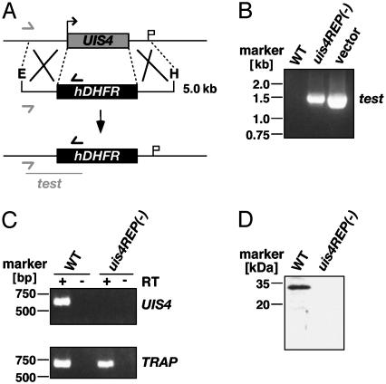
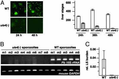
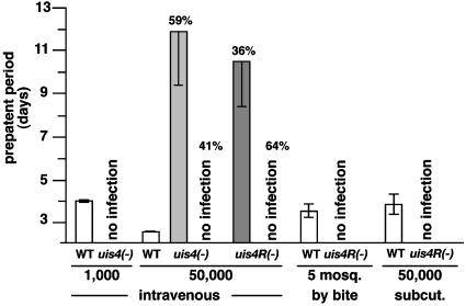
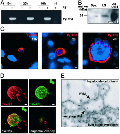
Plasmodium parasites of mammals, including the species that cause malaria in humans, infect the liver first and develop there into clinically silent liver stages. Liver stages grow and ultimately produce thousands of first-generation merozoites, which initiate the erythrocytic cycles causing malaria pathology. Here, we present a Plasmodium protein with a critical function for complete liver stage development. UIS4 (up-regulated in infective sporozoites gene 4) is expressed exclusively in infective sporozoites and developing liver stages, where it localizes to the parasitophorous vacuole membrane. Targeted gene disruption of UIS4 in the rodent model malaria parasite Plasmodium berghei generated knockout parasites that progress through the malaria life cycle until after hepatocyte invasion but are severely impaired in further liver stage development. Immunization with UIS4 knockout sporozoites completely protects mice against subsequent infectious WT sporozoite challenge. Genetically attenuated liver stages may thus induce immune responses, which inhibit subsequent infection of the liver with WT parasites.


Figures

Fig. 1.
Fig. 1.
Targeted gene disruption of PbUIS4 by a replacement strategy. (A) Replacement to generate the uis4REP- parasite. The WT UIS4 genomic locus is targeted with an EcoRI/HindIII-linearized replacement fragment containing 5′ and 3′ UTRs of the UIS4 ORF and the hDHFR-positive selectable marker. Upon a double crossover event, the UIS4 ORF is replaced by the positive selectable marker. Replacement-specific test primer combinations are indicated by arrows, and expected fragment is shown as lines. (B) Replacement-specific PCR analysis. Confirmation of the predicted gene-targeting in uis4REP- parasites is achieved by primer combinations that can only amplify from the recombinant locus. Black and gray arrows shown in A indicate primers that hybridize to regions in the plasmid backbone and outside the UIS4 ORF, respectively. (C) Depletion of UIS4 transcripts in uis4REP- parasites. cDNA from WT and uis4REP- sporozoites were amplified at 35 PCR cycles. UIS4 transcript is not detectable in knockout parasites when compared with a transcript control (TRAP). (D) Western blot analysis of WT and uis4REP- sporozoites. Extracts from 100,000 WT or uis4REP- salivary gland sporozoites were separated on a 10% SDS gel and probed with the polyclonal anti-UIS4 serum.
Fig. 2.
Fig. 2.
uis4- parasites arrest in liver stage development. (A) UIS4 is required for liver stage development in vitro.WTand uis4 - salivary gland sporozoites were added to subconfluent hepatoma cells, and the intracellular liver stages that develop were immunostained with anti-heat shock protein 70 at the indicated time points after infection. Shown in Right are mean liver stage numbers (±SEM) from four experiments each. (B) Late liver stages are not detected in mice infected with uis4- sporozoites. Five and eight mice were injected i.v. with 10,000 WT and uis4- sporozoites, respectively. After 48 h, livers were removed and subjected to total RNA extraction and RT-PCR. cDNA amplification was done with gene-specific primers for Pb 18S rRNA and mouse GAPDH, respectively. (C) Quantitative real-time RT-PCR confirms the absence of late liver stages in mice infected with uis4- sporozoites.
Fig. 3.
Fig. 3.
uis4- parasites are severely impaired in progression to blood stage infections in the mammalian host. WT and uis4- sporozoites were injected i.v. or s.c. or delivered by natural bite in numbers indicated. i.v. injection of 1,000 uis4- sporozoites (16 mice total) did not result in erythrocytic stage infections (patency), even after 21 days. WT-injected control mice (six mice total) become blood stage patent after 4 days (prepatent period). i.v. injection of 50,000 sporozoites resulted in delayed blood stage infections in uis4- and uis4REP- parasites at the proportion of animals indicated (56 and 75 mice total injected, respectively). Blood stage parasites from patent mice were genotyped and confirmed to be uis4- or uis4REP- mutants. Natural transmission by mosquito bite with five uis4REP- infected mosquitoes per mouse (10 mice total) or by s.c. injection of 50,000 uis4REP- sporozoites (10 mice total) did not result in blood stage infections. WT-infected control mice (10 mice total) became blood stage patent as indicated.
Fig. 4.
Fig. 4.
Plasmodium UIS4 is expressed throughout liver stage development and localizes to the parasite–host membrane interface. (A) PyUIS4 transcripts are present throughout in vivo liver stage development of Py. RT-PCR detects PyUIS4 expression in the liver of mice 10, 20, and 40 h after infection with 5 × 10⁵ sporozoites. No amplification is observed in control reactions without RT (-RT) and in an uninfected control mouse (ni). (B) Immunoblot analysis of Py salivary gland sporozoite extracts (Spz.) and extracts from Py-infected mouse livers at 44 h of liver stage development (LS). The right lane shows an extract of PyUIS4-Ad-infected HEK 293 cells (Ad-UIS4). (C) PyUIS4 is expressed in a circumferential pattern surrounding the parasite throughout liver stage development. Shown are Py liver stages in vivo at 12, 24, and 44 h of development. In all stages, PyUIS4 is localized around the parasite and in extensions that reach into the hepatocyte cytoplasm. (D) Three-dimensional serial reconstruction of a Pb liver stage (48 h) within a HepG2 cell from 25 confocal optical sections. The surface of the liver stage is visualized with anti-CSP staining, appearing uneven and patchy. PbUIS4 shows peripheral localization surrounding the liver stage and exhibits an irregular surface profile, with multiple vesicular protrusions, distinct when compared with the CSP profile. An extensive TVN is visible on one side of the liver stage, with multiple membranous vesicles and tubular structures reaching into the HepG2 cell cytoplasm, most of which maintain a connection to the PVM. Note that the CSP-positive growth-delayed small liver stage visible in the upper right corner of the micrographs does not show UIS4 expression. (Lower Right) Micrograph shows a1 μm, confocal optical section of the liver stage at a tangential plane. Vesicles of the TVN reveal a peripheral PbUIS4 localization most consistent with its localization in the vesicular membranes. (Scale bars: 5 μm.) (E) Immunoelectron microscopy confirms PyUIS4 localization to the PVM of liver stages in vivo. Five-nanometer gold particles decorate the PVM and a TVN protrusion that extends outward into the hepatocyte cytoplasm.

References

    1. Kappe, S. H., Kaiser, K. & Matuschewski, K. (2003) Trends Parasitol. 19, 135-143. - PubMed
    1. Meis, J. F., Verhave, J. P., Jap, P. H., Sinden, R. E. & Meuwissen, J. H. (1983) Nature 302, 424-426. - PubMed
    1. Meis, J. F., Verhave, J. P., Jap, P. H. & Meuwissen, J. H. (1985) Cell Tissue Res. 241, 353-360. - PubMed
    1. Meis, J. F. & Verhave, J. P. (1988) Adv. Parasitol. 27, 1-61. - PubMed
    1. Shortt, H. E. & Garnham, P. C. C. (1948) Nature 161, 126. - PubMed

Publication types

Substances

LinkOut - more resources