. 2005 Feb 15;102(7):2531-6.
doi: 10.1073/pnas.0409876102. Epub 2005 Feb 7.

Impaired vasodilation by red blood cells in sickle cell disease

Affiliations

Impaired vasodilation by red blood cells in sickle cell disease

John R Pawloski et al. Proc Natl Acad Sci U S A. .

Abstract

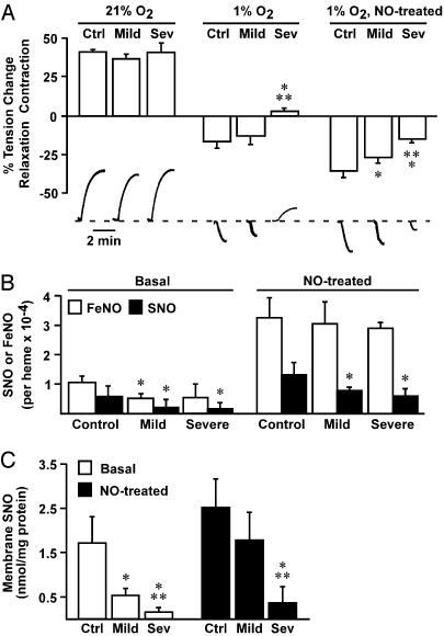
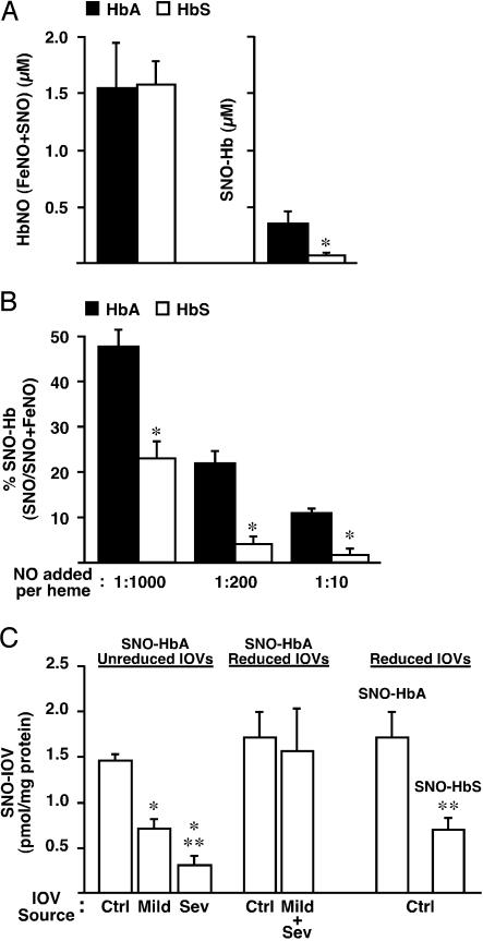
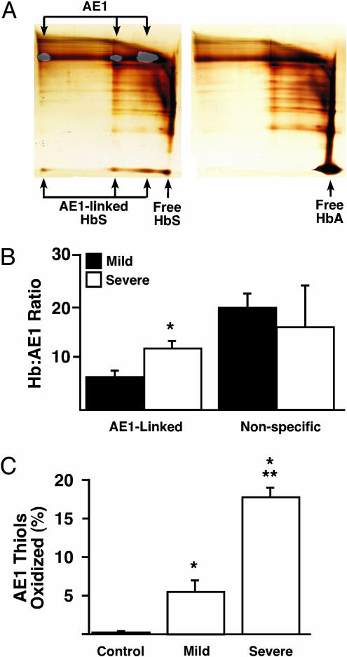
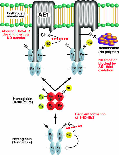
Red blood cells (RBCs) have been ascribed a unique role in dilating blood vessels, which requires O2-regulated binding and bioactivation of NO by Hb and transfer of NO equivalents to the RBC membrane. Vasoocclusion in hypoxic tissues is the hallmark of sickle cell anemia. Here we show that sickle cell Hb variant S (HbS) is deficient both in the intramolecular transfer of NO from heme iron (iron nitrosyl, FeNO) to cysteine thiol (S-nitrosothiol, SNO) that subserves bioactivation, and in transfer of the NO moiety from S-nitrosohemoglobin (SNO-HbS) to the RBC membrane. As a result, sickle RBCs are deficient in membrane SNO and impaired in their ability to mediate hypoxic vasodilation. Further, the magnitudes of these impairments correlate with the clinical severity of disease. Thus, our results suggest that abnormal RBC vasoactivity contributes to the vasoocclusive pathophysiology of sickle cell anemia, and that the phenotypic variation in expression of the sickle genotype may be explained, in part, by variable deficiency in RBC processing of NO. More generally, our findings raise the idea that defective NO processing may characterize a new class of hemoglobinopathy.


Figures

Fig. 1.
Fig. 1.
RBC vasoactivity and NO distribution in normal and sickle RBCs. (A) Oxygen-regulated vasoactivity in an aortic ring bioassay of RBCs from normal subjects (Ctrl) and from subjects with mild (Mild) or severe (Sev) SCD. All RBCs elicited vasoconstriction at 21% O2, consistent with scavenging of endothelial NO by R-state Hb. At tissue oxygen tension (1% O2, pO2 = 5–10 mmHg, favoring T-state Hb), normal and mild sickle RBCs evoked relaxation, but RBCs from severe disease subjects remained vasoconstrictive. Adjuvant NO amplified the hypoxic vasodilatory activity of control and mild RBCs, and transformed severe RBCs into vasodilators. Representative polygraph tracings of vasomotor activity are illustrated below. *, Significant with respect to control; **, significant with respect to mild SCD (n = 5–7 in each group; P < 0.05). (B and C) Both total SNO content (B) and membrane-associated SNO (C) were decreased in sickle vs. normal RBCs, but levels of membrane SNO differed significantly between mild and severe SCD and are thus more closely correlated with hypoxic vascular activity. Treatment with aqueous NO (1:250 NO/heme) increased and equalized FeNO in all groups, but total SNO content remained significantly lower in sickle vs. control RBCs (B), and membrane SNO content remained significantly lower in severe sickle RBCs (C). *, Significant with respect to control; **, significant with respect to mild SCD (n = 8–17 in each group; P < 0.05).
Fig. 2.
Fig. 2.
NO processing defects of sickle Hb and RBC membranes. (A) SNO-Hb formation by cell-free HbA and HbS (100 μM tetramer) after addition of aqueous NO (1:200 NO/heme). NO solution was added slowly to deoxygenated Hb followed by reoxygenation and filtration through Sephadex G-25. Quantitation of NO bound to heme (FeNO) and thiol (SNO) was carried out by photolysis-chemiluminescence. Total incorporation of NO (FeNO + SNO) was equivalent for HbA and HbS, but significantly less SNO-HbS was generated. *, Significant with respect to HbA (n = 4; P < 0.05). (B) SNO yield declines as the initial NO/heme ratio increases (absolute amount plateaus) (11), but the disparity between SNO formation by HbA vs. HbS is maintained. (C) Transfer of NO from immobilized SNO-HbA or SNO-HbS (generated from SNO-Cys and matched for SNO content) to IOVs prepared reductively (DTT/β-mercaptoethanol) or nonreductively from RBCs obtained from normal subjects (Ctrl) or from subjects with mild (Mild) or severe (Sev) SCD. Less NO was transferred from SNO-HbA to nonreduced sickle IOVs vs. nonreduced control IOVs (control > mild > severe), but transfer to control and sickle IOVs prepared reductively did not differ (Mild + Sev represents a mixture of IOVs from subjects with mild and severe disease). Note that NO group transfer from SNO-HbA to reduced vs. unreduced IOVs did not differ significantly. NO transfer from SNO-HbS to control IOVs (prepared reductively) was significantly decreased with respect to transfer from SNO-HbA. *, Significant with respect to nonreduced control IOV; **, significant with respect to nonreduced mild IOVs (left) or SNO-HbA (right) (n = 4–6 in each group; P < 0.05).
Fig. 3.
Fig. 3.
Hb cross-linking and AE1 thiol oxidation in native normal and sickle RBCs. (A) Two-dimensional gel analysis (nonreducing → reducing) of control (right) and sickle (left) RBC membranes. No disulfide-linked HbA was detected in association with membranes from normal RBCs. In contrast, HbS (lower arrows) was disulfide-linked to mono-, di-, and multimeric AE1 (upper arrows), identified by Western blot analysis of a gel run in parallel (low-intensity overlay). (Horizontal streaking represents dispersion in the first-dimension gel of cross-linked protein complexes, which did not differ between HbA and HbS.) (B) Association of HbS with sickle RBC membranes. The molar ratio of HbS disulfide-linked to AE1 was significantly greater in severe vs. mild SCD (11.19 vs. 6.34), whereas no difference was observed in the amount of membrane-associated but unlinked HbS. *, Significant with respect to mild SCD (n = 7–13 in each group; P < 0.05). (C) Analysis of AE1 thiol redox status, based on quantitation of free thiols with thiol-specific fluorescent labeling. AE1 thiol oxidation was more extensive in severe vs. mild sickle RBCs, whereas normal RBC AE1 thiols were essentially unoxidized. *, Significant with respect to normal; **, significant with respect to mild disease (n = 4–6 in each group; P < 0.05).
Fig. 4.
Fig. 4.
Schematic summary of the proposed defects in NO trafficking in the sickle erythrocyte, which inhibit generation of vasodilatory NO bioactivity at the RBC membrane. As shown at the bottom, the allosterically (O2/redox) regulated intramolecular transfer of NO from heme to thiol is impaired in HbS. This defect is likely to represent differences between HbS vs. HbA in heme redox potential (and in P50 upon polymerization), and thus in the ability to support S-nitrosylation. [Note that some portion of NO groups bound to β-heme will redistribute to α-heme (“recapture”) rather than thiol, that one oxygen is omitted from R-state SNO-Hb to provide for the possibility of β-heme oxidation (Fe2+ → Fe3+) coupled to SNO-Hb formation (6, 7), and that O2/redox-regulation of Hb is represented as a simplified two-state (R/T) model (6).] In addition, transfer of NO groups from SNO-HbS to a cysteine thiol within the CDAE1 is deficient, shown on the left, as a result of aberrant binding of HbS and CDAE1. As shown on the right, oxidative (disulfide) coupling of HbS to CDAE1, which may be facilitated by aberrant binding, results in the formation of cross-linked AE1-hemichrome complexes that preclude NO group transfer from SNO-HbS to the RBC membrane.

References

    1. McMahon, T. J., Moon, R. E., Luchsinger, B. P., Carraway, M. S., Stone, A. E., Stolp, B. W., Gow, A. J., Pawloski, J. R., Watke, P., Singel, D. J., et al. (2002) Nat. Med. 8, 711-717. - PubMed
    1. Pawloski, J. R., Swaminathan, R. V. & Stamler, J. S. (1998) Circulation 97, 263-267. - PubMed
    1. Pawloski, J. R., Hess, D. T. & Stamler, J. S. (2001) Nature 409, 622-626. - PubMed
    1. James, P. E., Lang, D., Tufnell-Barrett, T., Milsom, A. B. & Frenneaux, M. P. (2004) Circ. Res. 94, 976-983. - PubMed
    1. Datta, B., Tufnell-Barrett, T., Bleasdale, R. A., Jones, C. J., Beeton, I., Paul, V., Frenneaux, M. & James, P. (2004) Circulation 109, 1339-1342. - PubMed

Publication types