

A previously uncharacterized role for small protein B (SmpB) in transfer messenger RNA-mediated trans-translation
- PMID: 15699355
- PMCID: PMC549014
- DOI: 10.1073/pnas.0409694102
A previously uncharacterized role for small protein B (SmpB) in transfer messenger RNA-mediated trans-translation
Abstract
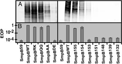
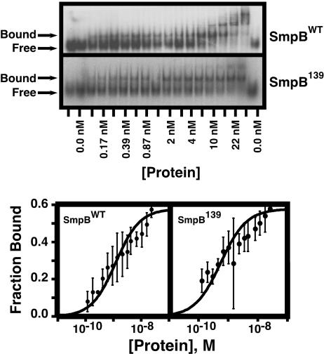
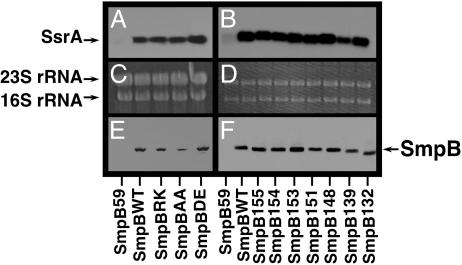
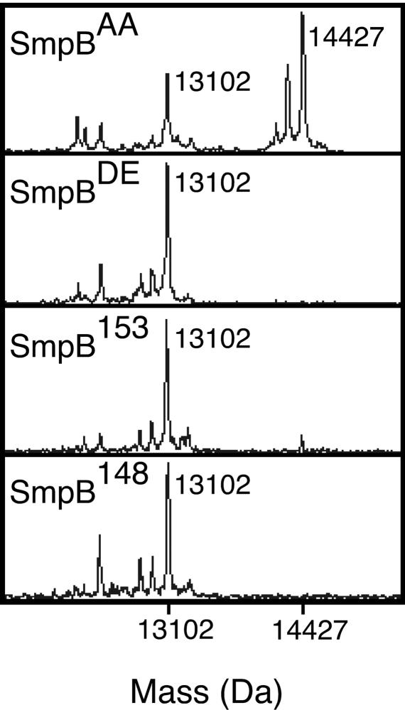
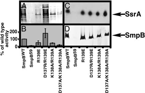
SsrA is a versatile RNA molecule found in all bacteria that functions as both a tRNA and an mRNA. SsrA rescues ribosomes stalled on damaged mRNAs and directs the tagging and degradation of their aberrant protein products. Small protein B (SmpB) is required for all known activities of SsrA. The two known functions of SmpB are binding SsrA RNA and promoting stable association of the SmpB.SsrA complex with 70S ribosomes. Using mutational analysis and biochemical experiments, we have discovered a previously uncharacterized SmpB function. This function is required for a step in the tagging process downstream of SsrA binding and ribosome association but before transpeptidation of the SsrA-linked alanine and establishment of the SsrA reading frame. Our results clearly demonstrate that residues in the C-terminal tail of SmpB confer a hitherto unrevealed function that is essential for trans-translation. Based on these results, we propose that upon binding stalled ribosomes, the unstructured C-terminal tail of SmpB acquires contacts that are critical for productive accommodation of SsrA into the ribosomal A site.
Figures
Similar articles
-
Crystal structure of the transfer-RNA domain of transfer-messenger RNA in complex with SmpB.Nature. 2003 Aug 7;424(6949):699-703. doi: 10.1038/nature01831. Nature. 2003. PMID: 12904796
-
SmpB, a unique RNA-binding protein essential for the peptide-tagging activity of SsrA (tmRNA).EMBO J. 1999 Jul 1;18(13):3793-9. doi: 10.1093/emboj/18.13.3793. EMBO J. 1999. PMID: 10393194 Free PMC article.
-
One SmpB molecule accompanies tmRNA during its passage through the ribosomes.FEBS Lett. 2008 Apr 30;582(10):1532-6. doi: 10.1016/j.febslet.2008.03.049. Epub 2008 Apr 7. FEBS Lett. 2008. PMID: 18396159
-
Dial tm for rescue: tmRNA engages ribosomes stalled on defective mRNAs.Curr Opin Struct Biol. 2004 Feb;14(1):58-65. doi: 10.1016/j.sbi.2004.01.010. Curr Opin Struct Biol. 2004. PMID: 15102450 Review.
-
The SsrA-SmpB system for protein tagging, directed degradation and ribosome rescue.Nat Struct Biol. 2000 Jun;7(6):449-55. doi: 10.1038/75843. Nat Struct Biol. 2000. PMID: 10881189 Review.
Cited by
-
Mechanisms of ribosome rescue in bacteria.Nat Rev Microbiol. 2015 May;13(5):285-97. doi: 10.1038/nrmicro3438. Epub 2015 Apr 13. Nat Rev Microbiol. 2015. PMID: 25874843 Review.
-
Lon protease degrades transfer-messenger RNA-tagged proteins.J Bacteriol. 2007 Sep;189(18):6564-71. doi: 10.1128/JB.00860-07. Epub 2007 Jul 6. J Bacteriol. 2007. PMID: 17616591 Free PMC article.
-
Small protein B interacts with the large and the small subunits of a stalled ribosome during trans-translation.Nucleic Acids Res. 2006 Apr 12;34(6):1935-43. doi: 10.1093/nar/gkl097. Print 2006. Nucleic Acids Res. 2006. PMID: 16611927 Free PMC article.
-
The highest affinity binding site of small protein B on transfer messenger RNA is outside the tRNA domain.RNA. 2008 Sep;14(9):1761-72. doi: 10.1261/rna.1185808. Epub 2008 Jul 22. RNA. 2008. PMID: 18648069 Free PMC article.
-
In vivo tmRNA protection by SmpB and pre-ribosome binding conformation in solution.RNA. 2014 Oct;20(10):1607-20. doi: 10.1261/rna.045674.114. Epub 2014 Aug 18. RNA. 2014. PMID: 25135523 Free PMC article.
References
-
- Keiler, K. C., Waller, P. R. & Sauer, R. T. (1996) Science 271, 990-993. - PubMed
-
- Gillet, R. & Felden, B. (2001) Mol. Microbiol. 42, 879-885. - PubMed
-
- Karzai, A. W., Roche, E. D. & Sauer, R. T. (2000) Nat. Struct. Biol. 7, 449-455. - PubMed
-
- Withey, J. H. & Friedman, D. I. (2003) Annu. Rev. Microbiol. 57, 101-123. - PubMed
Publication types
MeSH terms
Substances
LinkOut - more resources
Full Text Sources
Molecular Biology Databases
