. 2005 Feb 28;92(4):729-35.
doi: 10.1038/sj.bjc.6602385.

Early endostatin treatment inhibits metastatic seeding of murine colorectal cancer cells in the liver and their adhesion to endothelial cells

Affiliations

Early endostatin treatment inhibits metastatic seeding of murine colorectal cancer cells in the liver and their adhesion to endothelial cells

E A te Velde et al. Br J Cancer. .

Abstract

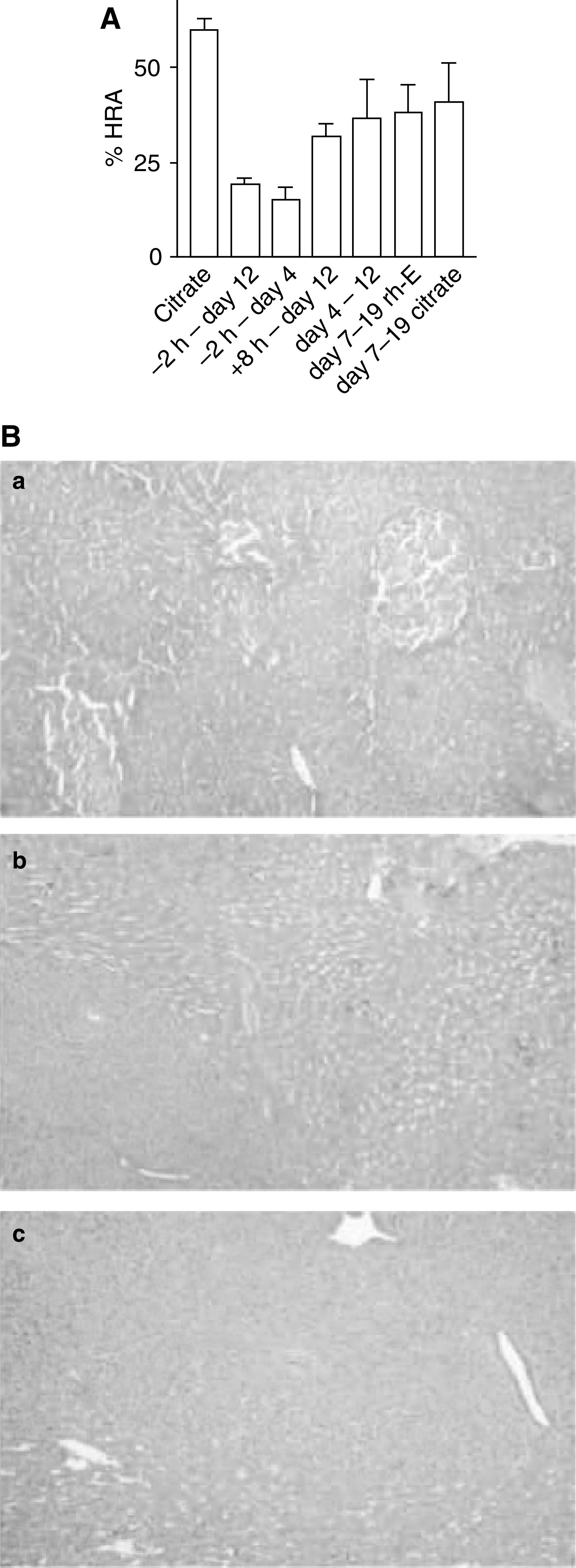
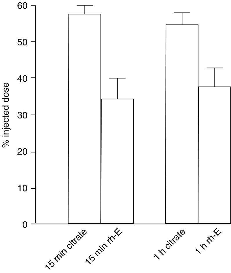
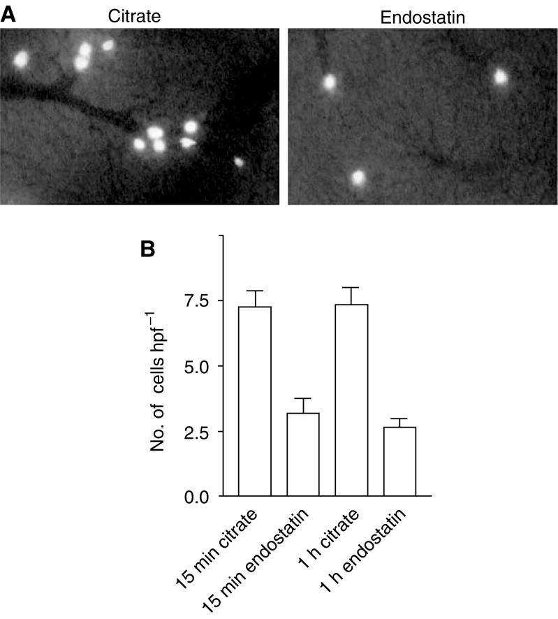
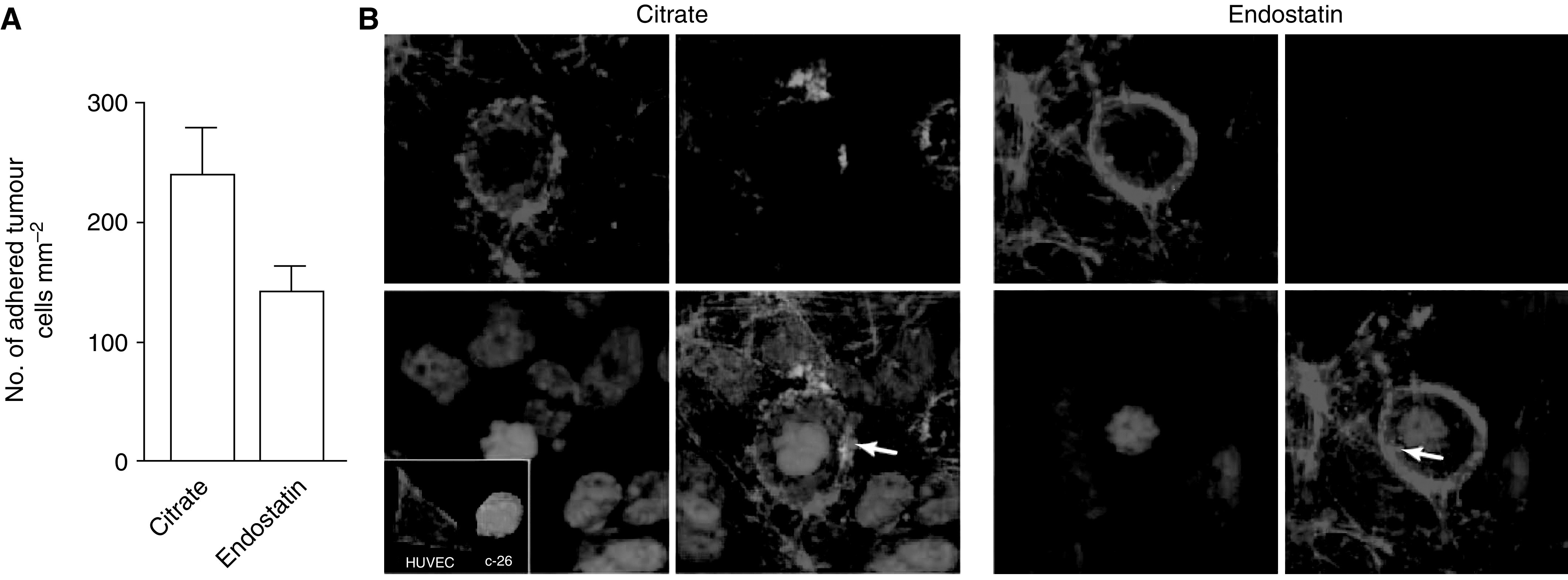
Endostatin, a carboxy-terminal fragment of collagen XVIII, potently inhibits angiogenesis and tumour growth, presumably through induction of apoptosis in endothelial cells and/or inhibition of their migration. Here we have tested how the timing of recombinant human endostatin (rh-E) administration affects its antitumour activity in a liver metastasis model of mouse C26 colorectal carcinoma cells. The effects of rh-E treatment on hepatic tumour load and on early tumour cell seeding were evaluated. Recombinant human endostatin was most effective in reducing intrahepatic tumour growth when administered prior to tumour cell inoculation. Analysis of early tumour cell seeding by using [(125)I]iododeoxyuridine-labelled C26 cells or by in vivo microscopy showed that rh-E reduced tumour cell seeding in the liver sinusoids. Recombinant human endostatin did not inhibit tumour growth when administered later than 4 days after tumour injection. Pretreatment of human umbilical vein endothelial cells with rh-E in vitro reduced C26 tumour cell adhesion under flow conditions two-fold as assessed by video microscopy and multiphoton laser scanning microscopy. Our results show that rh-E, in addition to antiangiogenic effects, reduces tumour cell adhesion in the liver sinusoids during the very early phases of metastasis formation. These data point towards a previously unknown mode of action of endostatin, that is, its ability to interfere with tumour cell seeding. Such insights may be helpful in the design of trials to improve (surgical) treatment of colorectal carcinoma and liver metastases.


Figures

Figure 1
Figure 1
(A) Endostatin (rh-E) therapy is most efficacious when started prior to tumour cell seeding. All mice were inoculated with tumour cells on t0. Recombinant human endostatin or control citrate buffer was given in different time schemes as indicated and the intrahepatic tumour load (HRA) for all different treatment groups was assessed on day 12. Data are plotted as means±s.e.m. (B) Reduced therapeutic efficacy of rh-E treatment on established tumours. (a) Haematoxylin- and eosin-stained sections from a liver 7 days after intrasplenic injection of tumour cells just prior to the start of treatment, showing three small intrahepatic tumour lesions (dark nodules). (b) Tumour-bearing liver 19 days after tumour cell injection treated with citrate buffer. (c) Tumour-bearing liver 19 days after tumour cell injection treated with rh-E. The tumours continued to grow under rh-E treatment. For quantification, see (A).
Figure 2
Figure 2
Radioactive labelled tumour cells in the liver, 15 min and 1 h after intrasplenic injection, as measured by gamma counter and represented as percentage of injected [125I]iododeoxyuridine-labelled tumour cells. At 15 min after tumour cell injection in the spleen, the percentage of injected dose in the liver was 34.4±5.6% in the mice that were treated with rh-E 2 h prior to tumour cell injection, vs 57.4±2.5% in the controls (P=0.001). After 1 h, these percentages had not significantly changed.
Figure 3
Figure 3
(A) Intravital microscopy images recorded 15 min after injection of fluorescent tumour cells into the spleen of a control liver and a liver after 2 h rh-E pretreatment. (B) Number of fluorescent tumour cells that are present in the liver per hpf as measured by intravital microscopy 15 min and 1 h after intrasplenic injection. Recombinant human endostatin reduced the number of arrested tumour cells in the liver by 56% (P<0.001). No differences were observed within either treatment group between the two time points (controls 15 min vs 1 h P=0.93; endostatin 15 min vs 1 h P=0.39).
Figure 4
Figure 4
Recombinant human endostatin reduces tumour cell adhesion under flow conditions. (A) C26 cells were allowed to adhere to a confluent layer of TNF-α-stimulated HUVECs under flow conditions for 5 min and this was recorded on a videotape. The number of adherent tumour cells per mm2 was determined by off-line analysis of the video images. Recombinant human endostatin reduced the number of adhered tumour cells to stimulated HUVECs when compared to citrate buffer control by 41% (P=0.04). (B) Immunofluorescence and multiphoton laser scanning microscopy visualised C26 tumour cell adhesion to HUVECs. After perfusion with tumour cells in full blood, the coverslips were fixed and stained with Hoechst (blue) to stain nuclei, Phalloidin-TRITC (red) to stain filamentous actin and anti-fibrinogen (green) to visualise the extracellular matrix. Platelets are indicated by arrows.

References

    1. Bergers G, Javaherian K, Lo KM, Folkman J, Hanahan D (1999) Effects of angiogenesis inhibitors on multistage carcinogenesis in mice. Science 284: 808–812 - PubMed
    1. Boehm T, Folkman J, Browder T, O'Reilly MS (1997) Antiangiogenic therapy of experimental cancer does not induce acquired drug resistance. Nature 390: 404–407 - PubMed
    1. Brandsma D, Reijeveld JC, Taphoorn MJ, de Boer HC, Gebbink MF, Ulfman LH, Zwaginga JJ, Voest EE (2002) Vascular cell adhesion molecule-1 is a key adhesion molecule in melanoma cell adhesion to the leptomeninges. Lab Invest 82: 1493–1502 - PubMed
    1. Chang Z, Choon A, Friedl A (1999) Endostatin binds to blood vessels in situ independent of heparan sulfate and does not compete for fibroblast growth factor-2 binding. Am J Pathol 155: 71–76 - PMC - PubMed
    1. Dixelius J, Cross M, Matsumoto T, Sasaki T, Timpl R, Claesson-Welsh L (2002) Endostatin regulates endothelial cell adhesion and cytoskeletal organization. Cancer Res 62: 1944–1947 - PubMed

Publication types