. 2005 Feb 8:5:14.
doi: 10.1186/1471-2407-5-14.

Quantitative evaluation and modeling of two-dimensional neovascular network complexity: the surface fractal dimension

Affiliations

Quantitative evaluation and modeling of two-dimensional neovascular network complexity: the surface fractal dimension

Fabio Grizzi et al. BMC Cancer. .

Abstract

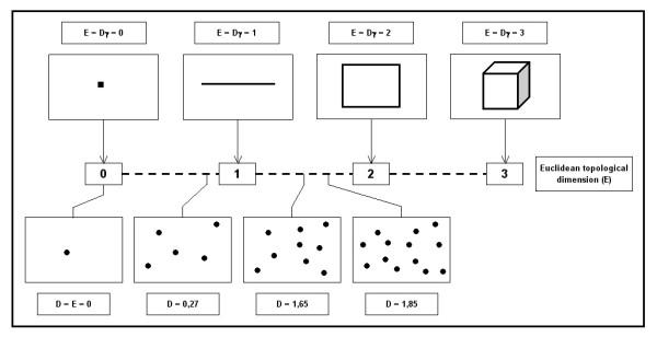
Background: Modeling the complex development and growth of tumor angiogenesis using mathematics and biological data is a burgeoning area of cancer research. Architectural complexity is the main feature of every anatomical system, including organs, tissues, cells and sub-cellular entities. The vascular system is a complex network whose geometrical characteristics cannot be properly defined using the principles of Euclidean geometry, which is only capable of interpreting regular and smooth objects that are almost impossible to find in Nature. However, fractal geometry is a more powerful means of quantifying the spatial complexity of real objects.

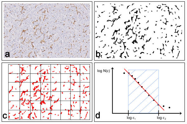
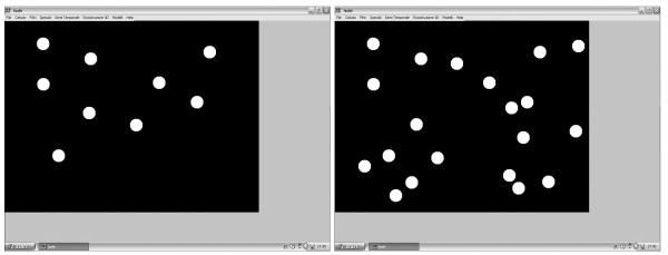
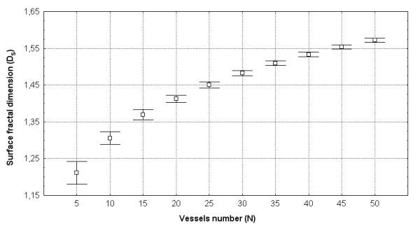
Methods: This paper introduces the surface fractal dimension (Ds) as a numerical index of the two-dimensional (2-D) geometrical complexity of tumor vascular networks, and their behavior during computer-simulated changes in vessel density and distribution.

Results: We show that Ds significantly depends on the number of vessels and their pattern of distribution. This demonstrates that the quantitative evaluation of the 2-D geometrical complexity of tumor vascular systems can be useful not only to measure its complex architecture, but also to model its development and growth.

Conclusions: Studying the fractal properties of neovascularity induces reflections upon the real significance of the complex form of branched anatomical structures, in an attempt to define more appropriate methods of describing them quantitatively. This knowledge can be used to predict the aggressiveness of malignant tumors and design compounds that can halt the process of angiogenesis and influence tumor growth.


Figures

Figure 1
Figure 1
Angiogenesis is a complex dynamic process that evolves through different states and a number of transitions between two successive states. At least seven critical steps have so far been identified in the sequence of angiogenic events on the basis of sprout formation.
Figure 2
Figure 2
The space-filling property of the vascular system is quantified by the fractal dimension (D), which falls between two topological integer dimensions. A. A Euclidean three-dimensional space (i.e. a cube) can contain a branching structure (i.e. the vascular system) without this entirely filling its internal space. B. Two-dimensional sectioning of the vascular network makes it possible to identify a variable number of vessels depending on the geometrical complexity of the system at any particular level of sectioning. C. The geometrical complexity of a 2-D section (s1, s2, s3) of the vascular network depends in the number of sectioned vessels and their distribution pattern.
Figure 3
Figure 3
Fractal dimensioning of the 2-D complexity of a vascular network. The figure shows four idealized cross-sectioned vascular patterns that not only have a different number of vessels, but also clearly different distributions: the geometrical complexity arising from these two variables determines the value of the surface fractal dimension.
Figure 4
Figure 4
Computer-aided estimate of the surface fractal dimension (Ds) of a vascular network in 2-D biopsy sections. A. Hepatocellular carcinoma section stained with antibodies raised against CD31 (Dako, Milan, Italy) that specifically react with vessels. B. Image segmentation: immunopositive vessels are specifically selected on the basis of the similarity of the color of adjacent pixels. C. Determination of Ds using the box-counting algorithm. Briefly, the method counts the number of boxes of length ε required to cover the object being measured, indicated as N(ε). D. Prototypical curve obtainable using the box-counting method that highlights the so-called fractal windows ranged by box size ε1 and ε2, and represents the appropriate region in which to estimate the dimension. Box sizes of more than ε2 approach the size of the image until one box covers it completely, at which point N(ε) = 1 and the slope = 0. Box sizes smaller than ε1 approach a single pixel or the resolution of the image: in this region, box counting simply gives the area of the image.
Figure 5
Figure 5
Computer-aided procedure used to quantify the surface fractal dimension of a simulated two-dimensional image of the vascular system. Prototypical 2-D simulated microscopy images of the vascular system with different vessel numbers and distribution were automatically generated, and their Ds was determined.
Figure 6
Figure 6
The behavior of Ds during a simulated increase in vessel density. The graph shows that different Dsvalues can be obtained for images with the same vessel density. As the only variable in these images is their distribution pattern, Ds depends on the irregular arrangement of the vessels in the surrounding environment (note the standard deviation of each cell density group). Ds also increases significantly when a higher vessel density is introduced into the system because of the greater space filled by the vascular component. The increase in vessel density reduces the variability of their space-filling properties, thus reducing the standard deviation.

References

    1. Carmeliet P. Angiogenesis in health and disease. Nature Medicine. 2003;9:653–670. doi: 10.1038/nm0603-653. - DOI - PubMed
    1. Woolf N. Pathology. Basic and Systemic. WB Saunders Company, London; 1998.
    1. Fidler IJ, Ellis LM. Neoplastic angiogenesis – not all blood vessels are created equal. New England Journal of Medicine. 2004;351:215–216. doi: 10.1056/NEJMp048080. - DOI - PubMed
    1. Fukumura D, Xavier R, Sugiura T, Chen Y, Park EC, Lu N, Selig M, Nielsen G, Taksir T, Jain RK, Seed B. Tumor induction of VEGF promoter activity in stromal cells. Cell. 1998;94:715–725. doi: 10.1016/S0092-8674(00)81731-6. - DOI - PubMed
    1. Tandle A, Blazer DG, Libutti SK. Antiangiogenic gene therapy of cancer: recent developments. Journal of Translational Medicine. 2004;2:22. doi: 10.1186/1479-5876-2-22. - DOI - PMC - PubMed

LinkOut - more resources